08 September 2020: Articles 
The Role of Ultrasound in Dunbar Syndrome: Lessons Based on a Case Report
Challenging differential diagnosis, Diagnostic / therapeutic accidents, Management of emergency care, Rare disease, Congenital defects / diseases, Educational Purpose (only if useful for a systematic review or synthesis)
Renato Farina- A Study design/planning
- B Data collection/entry
- C Data analysis/statistics
- D Data interpretation
- E Preparation of manuscript
- F Literature analysis/search
- *Corresponding author: [email protected]
- B Data collection/entry
- E Preparation of manuscript
- F Literature analysis/search
- B Data collection/entry
- E Preparation of manuscript
- F Literature analysis/search
- B Data collection/entry
- E Preparation of manuscript
- F Literature analysis/search
- B Data collection/entry
- E Preparation of manuscript
- F Literature analysis/search
- B Data collection/entry
- E Preparation of manuscript
- F Literature analysis/search
- B Data collection/entry
- E Preparation of manuscript
- B Data collection/entry
- E Preparation of manuscript
- B Data collection/entry
- E Preparation of manuscript
- B Data collection/entry
- E Preparation of manuscript
DOI: 10.12659/AJCR.926778
Am J Case Rep 2020; 21:e926778


Abstract
BACKGROUND: Dunbar syndrome is a rare vascular alteration caused by the abnormal course of the median arcuate ligament of the diaphragm, which in some patients causes chronic compression of the celiac artery and can cause non-specific symptoms such as diarrhea, retro-sternal pain, vomiting, swelling, and nausea, or a typical symptomatic triad with weight loss, post-prandial abdominal pain, and epigastric murmur. Color Doppler ultrasound and duplex Doppler ultrasound provide a complete diagnostic framework of this disease.
CASE REPORT: We describe a case of 55-year-old man with post-prandial epigastric pain, significant weight loss, and several episodes of retro-sternal pain. He underwent multidetector computed tomography of the abdomen and color duplex Doppler ultrasound examination of the celiac artery that highlighted stenosis of the celiac artery, more severe in expiratory apnea. The computed tomography showed the typical aspect of the celiac artery, with the “hook sign”. A duplex Doppler ultrasound examination showed a significant increase in peak speed (226 cm/s) due to severe stenosis of the celiac artery by the median arched ligament of the diaphragm.
CONCLUSIONS: This case is unique due to the severity of the celiac artery stenosis and the unusual clinical presentation of the patient who had frequent episodes of retro-sternal pain. The significant increase in peak velocity in the celiac artery in expiratory apnea, if associated with the typical symptomatology in the absence of other alterations of the splanchnic vessels, can be considered, in our opinion, sufficient for the diagnosis of Dunbar syndrome.
Keywords: Cardiovascular Abnormalities, Celiac Artery, multidetector computed tomography, Ultrasonography, Doppler, Color, Ultrasonography, Doppler, Duplex, Abdominal Pain, Constriction, Pathologic, median arcuate ligament syndrome, Ultrasonography
Background
Dunbar syndrome (DS) [1], also called median arcuate ligament syndrome (MALS) [2], is a rare vascular alteration similar to thoracic outlet syndrome [3], nutcracker syndrome [4], and May-Thurner syndrome [5], and is caused by the compression of an arterial or venous vessel by adjacent structures. In DS, the celiac artery (CA) and/or the surrounding neural ganglion are compressed by a tendon bundle of the diaphragm composed of the median arcuate ligament (MAL), which, together with the lateral arcuate ligament, form the lateral pillar of the lumbar portion of the diaphragm. The CA originates from the abdominal aorta (AA) at the level of the upper edge of the 1st lumbar vertebra, runs antero-inferiorly, passing under the MAL, and continuing antero-superiorly (Figure 1A, 1B). Ischemia and epigastric pain can increase with forced expiration, which causes the diaphragm to rise and the MAL to drop, further compressing the CA. Compression by the MAL can be asymptomatic thanks to the compensation of the numerous collateral shunts [6] between the CA and the superior mesenteric artery (Arc of Riolan, pancreatic-duodenal arcades, and Marginal artery of Drummond). However, these cases are not DS because to be considered such they must be symptomatic. However, the causes of this disease are still controversial and in fact, according to some authors, since the circulation that supplies the intraperitoneal viscera is very extensive, at least 2 significant stenoses must be present to have ischemia [7]. Some authors hypothesize that the typical pain of the syndrome is due to stenosis and ischemia of the CA [8], while others claim that the symptoms are caused by compression of the celiac plexus. In the literature, it is also claimed that there are congenital factors [9], but cases in which stenosis has occurred following surgery have been reported [10]. DS is most common in women ages 20–50 years. The typical symptomatology of DS is post-prandial epigastric pain and weight loss. The differential diagnosis must be based on imaging and clinical presentation, and must exclude pathologies that can cause the same symptomatology. Color Doppler ultrasound and duplex Doppler ultrasound (color Doppler US and duplex Doppler US) are the first-level examination for the diagnosis of DS, while the second-level examinations are computed tomography (CT) [11], selective angiography [12], and magnetic resonance imaging (MRI) [13]. DS is very rare: the incidence is estimated at around 2 for every 100 000 patients. The present case is unique in 2 ways. (1) The severity of the stenosis of the celiac artery in the absence of other vascular alterations. In the cases reported in the literature, stenosis of the CA was often associated with stenosis of the superior mesenteric artery or widespread atherosclerosis of the splanchnic vessels. (2) The unusual clinical presentation of the patient who, in addition to having typical post-prandial epigastric pain and weight loss, also had frequent episodes of retro-sternal pain in the absence of cardiac or gastric disease. Therapy consists of treatment with robotic or laparoscopic surgery with the release of the open ligament and celiac ganglionectomy [14,15] which causes regression of symptoms in 85% of patients. In cases of recurrence (7%), treatment with endovascular stenting may be indicated [16].
Case Report
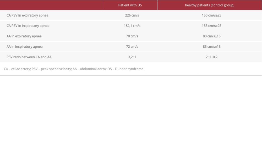
A 55-year-old man was sent to our hospital for observation after traffic accident trauma. The patient reported suffering from several years of post-prandial epigastric pain, having undergone a cholecystectomy for stones and in the last 5 months having had significant weight loss (11 kg in the past 6 months). After several episodes of retro-sternal pain, he underwent an endoscopic examination, which excluded gastro-duodenal alterations. He underwent a multidetector computed tomography (MDCT) angiography examination of the chest and abdomen. CT scans of the abdomen were performed in forced inspiration and forced expiration. An MDCT (Optima 64 slice, GE Healthcare) was used. The patient was also subjected to color and duplex Doppler US examination of the CA with an Aplio XG (Toshiba) device with a 3.5-MHz convex probe. Longitudinal and trans-verse scans were performed of the sub-xiphoid region with the patient in supine and lateral decubitus position, in inspiratory and expiratory apnea. The following measurements were performed in inspiratory and expiratory apnea: the maximum and minimum diameters, and the peak speed velocity (PSV) in stenotic, post-stenotic, and pre-stenotic tracts of the CA. The PSV ratio between AA and CA in expiratory apnea was calculated. The PSV of the AA was measured at the level of the CA origin. Doppler measurements were performed in 10 healthy patients (control group) and the results compared (Table 1). MDCT angiography excluded lesions of the abdominal and thoracic organs, and revealed a stenosis of the CA, more severe in expiratory apnea scans. In the reconstructions, according to a sagittal plane, it highlighted the “hook” appearance of the CA (Figure 2A–2C). The color Doppler US examination of the CA in expiratory apnea showed a turbulent flow and a minimum caliber of 3.3 mm in the stenotic tract, a 9-mm caliber in the post-stenotic tract, and an 11.5-mm caliber in the pre-stenotic tract. Ultrasound scans, performed in inspiratory apnea, showed a turbulent flow and a minimum caliber of 6.1 mm in the stenotic tract, a caliber of 8.6 in the post-stenotic tract, and caliber of 6.4 in the pre-stenotic tract (Figure 3A–3D).
PSV of CA in expiratory apnea in patient with DS was 226 cm/s and 182.1 cm/s in inspiratory apnea (Figure 4A, 4B; Video 1). The PSV ratio between CA and AA in expiratory apnea was 3.2: 1. In the symptom-free control group, PSV of CA in expiratory apnea was 150 cm/s±25 and the PSV ratio was 2: 1±0.2.
The same measurements were performed in the control group in 10 healthy patients. The results are summarized in the Table 1. The ultrasound study was performed by an operator with 20 years of experience in color Doppler US. The patient signed the informed consent form.
Discussion
Ultrasound is a first-level exam that plays an important role in the diagnosis of vascular compression syndromes, especially in patients with non-specific symptoms. An ultrasound examination limit is represented by the presence of excessive intestinal meteorism in the transverse colon that can prevent the optimal viewing celiac artery; in these cases, it may be useful to perform ultrasound scans with the patient in lateral decubitus position with support on the left side, which allows obtaining a good acoustic window for the study of the CA. To be affected by DS, patients must be symptomatic, otherwise it is only a congenital alteration of the celiac artery well-compensated flow rate. In our case, the diagnostic suspicion was cause by the patient’s clinical history. In fact, the post-prandial pain from continued even after the cholecystectomy and the onset of retro-sternal pain, which is unusual in this syndrome [17]; this, combined with the absence of gastro-duodenal alterations and weight loss, narrowed the diagnostic hypotheses. Furthermore, in an MRI examination performed before the cholecystectomy, a slight compression of the CA by the MAL was described. All these signs were decisive for diagnosis with the CT and US scans in inspiratory and expiratory apnea. In fact, the discovery of celiac artery stenosis was not accidental, despite the patient undergoing MDCT angiography only to exclude trauma of the abdominal organs. In many cases, the diagnosis of DS occurs incidentally, both because the patient has mild and non-specific symptoms and because ultrasound scans and MDCT angiography scans are generally performed in inspiratory apnea, and the stenosis may not be evident. Thus, if the symptoms are present, in the absence of other diseases, it is necessary, in our opinion to perform a diagnostic study with scans in expiratory apnea. In our case, B-mode US and color Doppler US allowed an accurate morphological study of the AA and AC, but the fundamental information for the diagnosis came from the flow measurements with the Doppler duplex US, which showed a significant increase of PSV with values greater than 200 cm/s in the expiratory apnea and a PSV ratio between the AC and the AA in the expiratory apnea 3.2: 1. In healthy patients, the PSV range between expiratory and inspiratory apnea varies between 150 cm/s and 155 cm/s±25 respectively). These findings, associated with the typical symptomatology, are sufficient for the diagnosis of DS. According to the literature, a PSV of the AC greater than 200 cm/s during the expiratory phase or a PSV ratio of the celiac artery to the aorta greater than 3: 1 in the expiratory phase are Doppler criteria for the diagnosis of DS. Some authors have described a borderline form of DS with only a slight increase in PSV (163.9 cm/s) without marked celiac compression in the US and CT [18]. The “hook sign”, shown by CT exam, is relatively common in daily practice, but in most cases it has no clinical significance. The CA impression is only an accessory sign that must be identified during a forced expiration.
Moreover, it is not always clearly identifiable in minor forms of DS since CT examinations are generally performed during the inspiratory phase. Surgery should only be considered in symptomatic patients and only after excluding all the other causes of abdominal pain. In our patient, due to the severity of the CA stenosis and according to the guidelines in the literature (Figure 5), the treatment we recommended was decompression, celiac ganglion sympathectomy, and selective revascularization with CA endovascular stenting (Figure 5) [19]. Traditional, laparoscopic or robotic surgical techniques [20] do not offer sufficient guarantees in the long term to avoid recurrence, and the combination of surgical treatment with AC stenting appears to be more effective. However, our patient refused treatment and after a few days he asked for and obtained discharge from the hospital.
Conclusions
Based on our experience, in patients with recurrent episodes of epigastric and retro-sternal pain, in absence of gastric and cardiological alterations, we recommend an ultrasound examination with color and duplex Doppler US to exclude DS. Ultrasound has numerous advantages compared to other imaging methods: it is a quick, well-tolerated, inexpensive, and repeatable examination and can be done while the patient is in bed; if performed by an experienced operator, can significantly reduce false negatives. We recommend a skeptical and cautious approach in choosing the appropriate therapy to avoid non-resolving interventions, choosing the intervention that offers greater long-term guarantees, which seems to us to be the hybrid technique: surgery + stenting. Failure to diagnose and treatment these patients could have serious implications for their quality of life.
Figures
References:
1.. Ali M, Patel J, Dunbar syndrome following liver transplantation: BMJ Case Rep, 2016; 2016; bcr2015214168
2.. Goodall R, Langridge B, Onida S, Median arcuate ligament syndrome: J Vasc Surg, 2020; 71(6); 2170-76
3.. Farina R, Foti PV, Iannace FA, Thoracic outlet syndrome: A rare case with bilateral cervical ribs and bilateral anterior scalene hypertrophy: J Ultrasound, 2019 [Online ahead of print]
4.. Farina R, Iannace FA, Foti PV, A Case of nutcracker syndrome combined with Wilkie syndrome with unusual clinical presentation: Am J Case Rep, 2020; 21; e922715
5.. Farina R, Foti PV, Iannace FA, May Thurner syndrome: Description of a case with unusual clinical onset: J Ultrasound, 2020 [Online ahead of print]
6.. Heo S, Kim HJ, Kim B, Clinical impact of collateral circulation in patients with median arcuate ligament syndrome: Diagn Interv Radiol, 2018; 24(4); 181-86
7.. Bech FR, Celiac artery compression syndromes: Surg Clin N Am, 1997; 77; 409-24
8.. Tribble CG, Harman PK, Mentzer RM, Celiac artery compression syndrome: Report of a case and review of the literature: Vasc Surg, 1986; 20; 120-29
9.. Bech F, Loesberg A, Rosenblum J, Median arcuate ligament compression syndrome in monozygotic twins: J Vasc Surg, 1994; 19(5); 934-38
10.. Takeda FR, Darce GFB, Sobrado LF, Post-esophagectomy symptomatic Dunbar syndrome: A rare diagnosis of abdominal pain after surgery: Int J Surg Case Rep, 2020; 68; 198-202
11.. Baskan O, Kaya E, Gungoren FZ, Erol C, Compression of the celiac artery by the median arcuate ligament: Multidetector computed tomography findings and characteristics: Can Assoc Radiol J, 2015; 66(3); 272-76
12.. Santos GM, Viarengo LMA, Oliveira MDP, Celiac artery compression: Dunbar syndrome: J Vasc Bras, 2019; 18; e20180094
13.. Aschenbach R, Basche S, Vogl TJ, Compression of the celiac trunk caused by median arcuate ligament in children and adolescent subjects: Evaluation with contrast-enhanced MR angiography and comparison with Doppler US evaluation: J Vasc Interv Radiol, 2011; 22(4); 556-61
14.. Roberts B, Pevsner R, Alkhoury F, Robotic approach for median arcuate ligament release in pediatrics: J Laparoendosc Adv Surg Tech A, 2020; 30(1); 92-96
15.. San Norberto EM, Romero A, Fidalgo-Domingos LA, Laparoscopic treatment of median arcuate ligament syndrome: A systematic review: Int Angiol, 2019; 38(6); 474-83
16.. Hongsakul K, Rookkapan S, Sungsiri J, Tubtawee T, A severe case of median arcuate ligament syndrome with successful angioplasty and stenting: Case Rep Vasc Med, 2012; 2012; 129870
17.. Karavelioğlu Y, Kalçık M, Sarak T, Dunbar syndrome as an unusual cause of exercise-induced retrosternal pain: Turk Kardiyol Dern Ars, 2015; 43(5); 465-67
18.. Acampora C, Di Serafino M, Iacobellis F, Insight into Dunbar syndrome: color-Doppler ultrasound findings and literature review: J Ultrasound, 2020 [Online ahead of print]
19.. Michalik M, Dowgiałło-Wnukiewicz N, Lech P, Hybrid (laparoscopy + stent) treatment of celiac trunk compression syndrome (Dunbar syndrome, median arcuate ligament syndrome (MALS)): Wideochir Inne Tech Maloinwazyjne, 2016; 11(4); 236-39
20.. You JS, Cooper M, Nishida S, Treatment of median arcuate ligament syndrome via traditional and robotic techniques: Hawaii J Med Public Health, 2013; 72(8); 279-81
Figures
In Press
16 Mar 2024 : Case report ")
Am J Case Rep In Press; DOI: 10.12659/AJCR.943214
16 Mar 2024 : Case report 
Am J Case Rep In Press; DOI: 10.12659/AJCR.943010
16 Mar 2024 : Case report 
Am J Case Rep In Press; DOI: 10.12659/AJCR.943687
17 Mar 2024 : Case report 
Am J Case Rep In Press; DOI: 10.12659/AJCR.943070
Most Viewed Current Articles
07 Mar 2024 : Case report 
DOI :10.12659/AJCR.943133
Am J Case Rep 2024; 25:e943133
10 Jan 2022 : Case report 
DOI :10.12659/AJCR.935263
Am J Case Rep 2022; 23:e935263
19 Jul 2022 : Case report 
DOI :10.12659/AJCR.936128
Am J Case Rep 2022; 23:e936128
23 Feb 2022 : Case report
DOI :10.12659/AJCR.935250
Am J Case Rep 2022; 23:e935250