29 July 2022: Articles 
Systemic Brucellosis with Arrhythmogenic Cardiac Inflammatory Pseudotumor
Challenging differential diagnosis, Unusual or unexpected effect of treatment, Rare coexistence of disease or pathology
Krzysztof KaczmarekDOI: 10.12659/AJCR.935259
Am J Case Rep 2022; 23:e935259


Abstract
BACKGROUND: Cardiac inflammatory pseudotumors are rarely observed. Their etiology might include immunologic abnormalities, fibrogenetic disorders, specific reactions to infections or abnormalities related to trauma, necrosis, or neoplasm. Life-threatening ventricular tachycardia and cases of sudden death related to cardiac tumors have been reported. The present report describes and discusses diagnostic and therapeutic solutions for the treatment of nonsarcoid multiorgan pseudotumors with cardiac involvement.
CASE REPORT: A 38-year-old woman presented to the clinic with symptomatic ventricular tachycardia. As coronary artery disease, cardiomyopathy, and channelopathy were ruled out, and electrocardiograms were not typical of idiopathic arrhythmia, the patient underwent detailed diagnostics which included targeted endomyocardial biopsy, which revealed a cardiac inflammatory pseudotumor. Laborious testing (and eventually, antibiotic therapy) led to ex juvantibus diagnosis of multiorgan disseminated brucellosis with cardiac involvement. Treatment with ceftriaxone, doxycycline, and rifampicin resulted in a complete resolution of all lesions after 3 months, and sustained recovery was observed during a 5-year follow-up. As the risk of ventricular tachycardia could not be reliably predicted, the patient had a subcutaneous implantable cardioverter-defibrillator implanted.
CONCLUSIONS: A vast diagnostic armamentarium of modern medicine allowed us to diagnose an unsuspected and rare cardiac inflammatory pseudotumor. In the case of travelers, the possibility of regionally specific illnesses, especially infections, must be taken into consideration as possible causes of arrhythmias. Cardiac magnetic resonance imaging may be useful in patients with ‘idiopathic ventricular tachycardias’ to detect non-apparent myocardial lesions which may result from the underlying cause of the arrhythmia.
Keywords: brucellosis, case reports, Tachycardia, Ventricular, Adult, Arrhythmias, Cardiac, Defibrillators, Implantable, Female, Granuloma, Plasma Cell, Heart Neoplasms, Humans
Background
The initial assessment of an episode of sustained ventricular tachycardia (VT) typically includes 12-lead ECG, transthoracic echocardiography, and functional testing and/or imaging for coronary artery abnormalities [1]. If structural or electrical heart disease is not excluded, a patient is deemed to have idiopathic VT, and further treatment, either pharmacological or invasive, must be planned, with consideration for the patient’s preference [2]. However, in rare cases, a thorough diagnostic process may lead to unsuspected findings. Infections should also be taken into consideration as possible causes of arrhythmias. Isolated cases of arrhythmia-triggering cardiac pseudotumors have been reported. Their etiology might include immunologic abnormalities, fibrogenetic disorders, specific reactions to infections, or abnormalities related to trauma, necrosis, or neoplasm. In the case of travelers, the possibility of contracting regionally specific illnesses, especially infections (including zoonoses), must be taken into consideration as possible causes of arrhythmias.
Case Report
A 38-year-old woman, with no previous medical history, presented in our hospital with palpitations which had lasted a few hours and were associated with 2 episodes of presyncope. Ventricular tachycardia of approximately 180 bpm (Figure 1) was diagnosed and sinus rhythm was restored with direct-current cardioversion 200J (Schiller DEFIGARD 5000).
Ventricular tachycardia recurred after several days and required another cardioversion. Initial evaluation included basic laboratory blood tests, the results of which were found to be normal, and transthoracic echocardiography that disclosed no abnormality. ECG revealed sinus bradycardia, lack of R-wave progression V1-4, and non-specific repolarization abnormalities with QTc falling within the normal range (QTc=380 ms) (Figure 2). The patient presented adequate exercise tolerance, heart rate, and blood pressure reactions, with no ECG changes during the treadmill test. Coronary angiography disclosed no abnormalities. The patient denied having any history of ventricular arrythmia events, sudden cardiac deaths, or premature deaths in her relatives. As typical pathologies related to VT (coronary artery disease, cardiomyopathies, and channelopathies) were excluded, idiopathic VT was considered as a primary working diagnosis. However, an electrophysiologist opined that our patient’s ECGs did not match a typical picture of idiopathic VT and scar-related arrhythmia was suspected (detailed description in Figure 1 footnote). Therefore, the patient was referred for cMRI, which revealed tumorous contrast enhancement in the middle portion of interventricular septum (1×2.7 cm) and in the subendocardial region, in the basal segment of the right ventricle (Figure 3).
As the nature of the cardiac tumor could not be precisely established based on cMRI, a whole-body positron emission tomography scan (18F-FDG-PET/CT) was performed. The scan revealed several foci of increased metabolism in multiple organs with maximal standardized uptake value (SUVmax) varying from 5.3 to 14.5 (Figure 4).
Additionally, MRI of the brain disclosed a 12×13×10 mm lesion in the central part of the pons (Figure 5). The patient presented no symptoms typical of localized or disseminated infection or myocarditis. The markers for systemic inflammation and myocardial injury were within normal ranges. Disseminated sarcoidosis was considered as the most probable initial diagnosis. Transbronchial biopsy of pulmonary foci, however, appeared to be inconclusive as no abnormal cells were found on histopathology. For the abdominal foci, it was too risky to undergo biopsy, so targeted biopsy of the septal focus in the heart was performed. For this purpose, we used multiple-imaging guidance, which included intraoperative fluoroscopy, transesophageal echocardiography (TEE), and 3D electrophysiological mapping with the CARTO System (Biosense Webster Inc., Irvine, CA, USA) (Figure 6).
The echocardiographer led the bioptome to the middle of the septum and electroanatomical mapping showed areas of subtle reduction of voltage which were considered to be connected with the tumor. The subsequent histopathology disclosed an inflammatory pseudotumor (IP) with no signs of sarcoidosis (Figure 7).
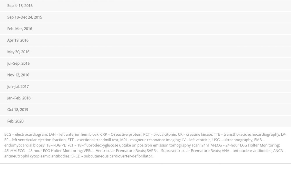
Therefore, bearing in mind that the patient had visited 4 different continents during the previous 6 months, stepwise diagnostics (Table 1) of possible causes of multiorgan IP were undertaken. The following tests gave positive results: (1) IgG antibodies against
Multiorgan brucellosis or an autoimmunological disorder did not seem likely, but they were not impossible either. However, having considered a possibility of infection with
Discussion
We describe here the case of a patient in whom multiorgan brucellosis with cardiac involvement in the form of intraventricular tumor provoking symptomatic VT was considered the most probable diagnosis.
The possibility that the lesion was a neoplastic tumor was the keynote of our decisions; therefore, a whole-body PET/CT was performed. SUVmax of the foci fell within the range of overlap between malignant neoplasms and inflammatory lesions [3]. As the biopsy of foci in peripheral organs was either unsuccessful or posed too high a risk to be performed, targeted transvenous biopsy of the interventricular septum was performed. Although a combination of echocardiography and fluoroscopy is usually used to guide biopsies of heart tumors [4], we could not apply this technique because our patient’s tumor was not clearly visible in echocardiography. Therefore, we performed additional 3D electroanatomical reconstruction of the intraventricular septum, which is known to target myocardial scars in cardiomyopathies and cardiac masses [5]. Our approach was unique in such a setting because in the majority of the reported cases of inflammatory cardiac pseudotumors, histopathology was performed either as post-mortem autopsy or after cardiac surgeries that often entailed severe or lethal complications [6].
The term “inflammatory pseudotumors” describes various inflammatory masses that are known by different names (most commonly as inflammatory myofibroblastic tumors, but also as plasma cell granuloma, histiocytoma, and others). IP etiology might include immunologic abnormalities, fibrogenetic disorders, specific reactions to an infectious agent, or abnormalities related to trauma, necrosis, or neoplasm. The clinical course of IPs has not been precisely determined and their classification is still uncertain. To date, neither diagnostic schemes nor adequate treatments have been agreed on [6–8]. IP secondary to infections, including brucellosis, have only rarely been described.
As the biopsy revealed neither signs of neoplasm nor of sarcoidosis, a differential diagnosis became even more challenging because the underlying causes of non-sarcoid inflammatory pseudotumors include a variety of pathologies that are not commonly diagnosed by cardiologists. Therefore, after multi-disciplinary consultations, diagnostic tests for specific infections (bacterial, viral, fungal, protozoal, and helminthic) as well as rheumatic diseases were performed (Table 1). The obtained results were not straightforward, but did narrow down the probable causes of the underlying pathologies to 2 possibilities: brucellosis or an autoimmune disorder. Anti-inflammatory therapy with steroids could potentially exacerbate the course of infection if the cause were indeed brucellosis, and it was for this reason that we decided to initiate antimicrobial treatment first. As this resulted in full resolution of the inflammatory lesions, the final diagnosis of the underlying cause of the IP was
To the best of our knowledge, only one case of an inflammatory tumor in the interventricular septum of the heart has been published, and its underlying cause was not disclosed [9]. Cardiac involvement during
Life-threatening VTs and cardiac tumor-related sudden death cases have been reported [13]. The future risk of VT recurrence could not be reliably estimated in our patient due to the extremely rare etiology and uncertain dynamics of her inflammatory cardiac lesions. As no stratification method (ie, invasive electrophysiological study) has been demonstrated in such a clinical scenario, we decided to implant a cardioverter-defibrillator. As the patient did not require permanent pacing and, bearing in mind that over the course of a 3-month in-hospital observation she had no VT recurrence, a subcutaneous device was chosen instead of a transvenous system. Disadvantages of S-ICD systems include the inability to provide antitachycardia pacing in case of monomorphic VT; however, by not implanting the defibrillation lead into the right ventricle, we avoided its possible harmful interaction with the injured myocardium [14]. Other possible options could be a wearable cardioverter-defibrillator but taking into account observations of recurrences of brucellosis, we decided that permanent protection with the implantable device might be a better option.
Conclusions
A vast diagnostic armamentarium of modern medicine allowed us to diagnose an unsuspected and rare cardiac inflammatory pseudotumor. In the case of travelers, the possibility of regionally specific illnesses, especially infections, must be taken into consideration as possible causes of arrhythmias. Cardiac magnetic resonance imaging may be useful in patients with ‘idiopathic ventricular tachycardias’ to detect non-apparent myocardial lesions, which may prove to be an underlying cause of the arrhythmia. Even though diagnosis
Figures
References:
1.. Priori SG, Blomström-Lundqvist C, Mazzanti A, 2015 ESC Guidelines for the management of patients with ventricular arrhythmias and the prevention of sudden cardiac death: The Task Force for the Management of Patients with Ventricular Arrhythmias and the Prevention of Sudden Cardiac Death of the European Society of Cardiology (ESC). Endorsed by: Association for European Paediatric and Congenital Cardiology (AEPC): Eur Heart J, 2015; 36; 2793-867
2.. Xiong Y, Zhu H, Electrocardiographic characteristics of idiopathic ventricular arrhythmias based on anatomy: Ann Noninvasive Electrocardiol, 2020; 25; e12782
3.. Meng J, Zhao H, Liu Y, Assessment of cardiac tumors by 18F-FDG PET/ CT imaging: Histological correlation and clinical outcomes: J Nucl Cardiol, 2021; 28(5); 2233-43
4.. Zanobini M, Dello Russo A, Saccocci M, Endomyocardial biopsy guided by intracardiac echocardiography as a key step in intracardiac mass diagnosis: BMC Cardiovasc Disord, 2018; 18; 1-5
5.. Burrell LD, Weiss PJ, Whisenant BK, Biopsy of a complicated right atrial mass using CARTO 3-dimensional electro-anatomic mapping: Catheter Cardiovasc Interv, 2014; 84; E61-64
6.. Kato T, Tomita S, Tamaki M, Inflammatory myofibroblastic tumor of the heart: Heart Vessels, 2014; 29; 123-28
7.. Burke A, Tavora F, The 2015 WHO Classification of tumors of the heart and pericardium: J Thorac Oncol, 2016; 11; 441-52
8.. Karnik A, Awtry E, Management of inflammatory cardiac masses: Cardiol Res, 2018; 9; 400-6
9.. Deng MD, Han JY, Lin K, Tang H, Cardiac inflammatory pseudotumor in inter-ventricular septum: A rare case report: Medicine (Baltimore), 2018; 96; e13219
10.. Pappas G, Akritidis N, Bosilkovski M, Tsianos E, Brucellosis: N Engl J Med, 2005; 352; 2325-36
11.. Vassiliadis T, Vougiouklis N, Patsiaoura K, Inflammatory pseudotumor of the liver successfully treated with nonsteroidal anti-inflammatory drugs: A challenge diagnosis for one not so rare entity: Eur J Gastroenterol Hepatol, 2007; 19; 1016-20
12.. McCarthy M, Anderson E, Debrito P, Successful treatment of recurrent inflammatory pseudotumor with high-dose prednisone: J Bronchol Interv Pulmonol, 2018; 25; e52-54
13.. Kusano KF, Ohe T, Cardiac tumors that cause arrhythmias: Card Electrophysiol Rev, 2002; 6; 174-77
14.. Steffel J, The subcutaneous ICD for prevention of sudden cardiac death: Current evidence and future directions: Pacing Clin Electrophysiol, 2020; 43; 1421-27
15.. Norbis L, Miotto P, Alagna R, Cirillo DM, Tuberculosis: Lights and shadows in the current diagnostic landscape: New Microbiol, 2013; 36; 111-20
Figures
In Press
Case report 
Am J Case Rep In Press; DOI: 10.12659/AJCR.953173
Case report 
Am J Case Rep In Press; DOI: 10.12659/AJCR.953192
Case report 
Am J Case Rep In Press; DOI: 10.12659/AJCR.952818
Case report
Am J Case Rep In Press; DOI: 10.12659/AJCR.953608
Most Viewed Current Articles
07 Dec 2021 : Case report
22,364,578
DOI :10.12659/AJCR.934347
Am J Case Rep 2021; 22:e934347
06 Dec 2021 : Case report  174,245
DOI :10.12659/AJCR.934406
Am J Case Rep 2021; 22:e934406
21 Jun 2024 : Case report
119,744
DOI :10.12659/AJCR.944371
Am J Case Rep 2024; 25:e944371
07 Mar 2024 : Case report
64,648
DOI :10.12659/AJCR.943133
Am J Case Rep 2024; 25:e943133