Logo American Journal of Case Reports

Call: 1.631.629.4328
Mon-Fri 10 am - 2 pm EST

Contact Us

Logo American Journal of Case Reports Logo American Journal of Case Reports Logo American Journal of Case Reports

20 October 2023: Articles  Japan

Infection Presenting with Myalgia and Cellulitis

Unusual clinical course, Challenging differential diagnosis

Hitomi Shimada12AEF, Risa Hirata ORCID logo1AEF, Shun Yamashita ORCID logo1E, Yuka Hirakawa1E, Orisa Nakashima1E, Masahiko Nakamura1E, Shizuka Yaita1E, Midori Tokushima1E, Yoshinori Tokushima1E, Hidetoshi Aihara1E, Motoshi Fujiwara1E, Masaki Tago ORCID logo1AE*

DOI: 10.12659/AJCR.941777

Am J Case Rep 2023; 24:e941777

0 Comments

Abstract

0:00

BACKGROUND: Helicobacter cinaedi is a rare bacterium, accounting for only 0.2% of the positive isolates in blood cultures. Previous reports note that patients with H. cinaedi infection often have underlying diseases. H. cinaedi infection is diagnosed by blood culture. However, because of the slow growth of this bacterium in blood culture, the diagnosis can be missed.

CASE REPORT: A 78-year-old man gradually developed erythema and pain in his left arm, then left shoulder and both lower legs. The patient presented to our hospital on day 17. He was afebrile, but the examination was remarkable for tenderness in both gastrocnemius muscles and erythema from the distal left lower leg to the ankle. We suspected pyomyositis and cellulitis and started oral administration of amoxicillin-clavulanate. On day 22, H. cinaedi was detected in blood cultures. Based on these findings, we diagnosed pyogenic myositis and cellulitis caused by H. cinaedi bacteremia. On day 24, antibiotic therapy was changed to intravenous ampicillin, and symptoms improved. Additional examination did not reveal any underlying immunodeficiency disorder, such as malignancy or HIV infection.

CONCLUSIONS: H. cinaedi infection can occur in healthy patients. Myalgia can be caused by pyogenic myositis because of bacteremia. In cases of myalgia or cellulitis of unknown etiology, blood cultures can be useful when bacteremia is suspected; blood samples should be monitored over an extended period.

Keywords: Bacteremia, Blood Culture, Helicobacter cinaedi, Myalgia, Male, Humans, Aged, Cellulitis, HIV Infections, Erythema, myositis

Background

Helicobacter cinaedi is a gram-negative, spiral-shaped bacterium. It is an intestinal resident, first isolated from the rectum of a homosexual man in the United States in 1984 [1]. The possibility of bacterial translocation from the intestinal tract has been suggested as a route of infection [2]. Previous reports have described cases of H. cinaedi infection in immunocompromised patients with underlying conditions such as malignancy, acquired immune deficiency syndrome, and chronic kidney disease (treated with hemodialysis) [3,4]. In recent years, there have also been reports of H. cinaedi infection among healthy individuals in the community [5]. Although the symptoms of H. cinaedi infection are nonspecific, fever can be the only symptom [6], and other symptoms such as cellulitis, gastroenteritis, enteritis, erysipelas, and arthritis have been reported. H. cinaedi infection is usually diagnosed by the detection of H. cinaedi in blood cultures, but it can be overlooked because the bacterial growth is slow and its morphology is similar to that of other species [3]. In addition, there has been a report of a recurrent case of H. cinaedi infection in which appropriate treatment was not implemented for several months because of a failure to diagnose the infection, resulting in repeated episodes of fever and rash [7]. An accurate diagnosis and appropriate treatment are crucial. We herein report a case of H. cinaedi infection in a non-immunocompromised individual who presented with myalgia and cellulitis and was subsequently diagnosed by blood cultures.

Case Report

A healthy 78-year-old man developed erythema and warmth in the left elbow to the left forearm and both distal lower legs on day 0. The patient had no history of pet ownership, trauma, or sexual activity. On day 1, he visited his primary care physician, and he had inflammation in the left elbow joint; aspiration of the joint revealed clear fluid. The patient was prescribed nonsteroidal anti-inflammatory drugs, which improved the erythema in the left forearm, although the symptoms in both distal lower legs persisted. The patient developed sharp and severe sharp pain in the left shoulder on day 6, and stabbing pain in the gastrocnemius muscles of both legs and the left lower leg on day 7. Finally, on day 17, he presented to our hospital. During the clinical course, the patient had no symptoms such as diarrhea or abdominal pain suggesting gastrointestinal infection.

On admission, his vital signs were body temperature, 35.5°C; blood pressure, 150/80 mmHg; heart rate, 102 beats/min; respiratory rate, 12 breaths/min; and percutaneous oxygen saturation, 98% (room air). His body mass index was 21.5 kg/m2. Physical examination revealed tenderness in the gastrocnemius muscles of both legs and erythema in the left lower leg to the ankle area, but there was no swelling or warmth. There were no other remarkable findings on physical examination of the limbs, and no rash was observed. Physical examination of the chest and abdomen were also unremarkable.

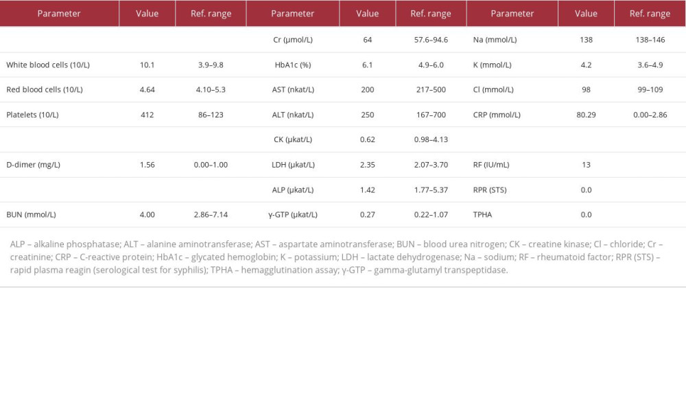
Blood tests showed a white blood cell count of 10.1×10⁹/L, with neutrophils 69.6% (reference value: 41.8–73.8%), lymphocytes 21.5% (18.3–47.5%), and no abnormal cells in the peripheral blood. Hemoglobin level was 141 g/L, and platelet count was 412×109/L. Kidney and liver function were normal, with lactate dehydrogenase (LDH) level 2.35 µkat/L, C-reactive protein (CRP) level 80.29 mmol/L, ferritin level 0.611 nmol/L (0.04–0.562 nmol/L), soluble interleukin 2 receptor (sIL-2R) level 749 U/mL (121–613 U/mL), carcinoembryonic antigen (CEA) level 2.5 ng/mL (<5.0 ng/mL), and carbohydrate antigen 19-9 (CA19-9) level 11 U/mL (<37 U/mL). Syphilis and HIV antibodies were negative, and there was no complement deficiency. Anti-nuclear antibody, anti-aminoacyl tRNA synthetase (ARS) antibody, and anti-neutrophil cytoplasmic antibodies (ANCA) were negative as well (Table 1). At the first visit to our hospital on day 17, blood cultures using the BACTEC system were obtained. Urine tests were normal. Contrast-enhanced computed tomography of the chest, abdomen, and lower limbs revealed soft tissue swelling and subcutaneous fat tissue opacity in the left lower leg, extending to the ankle (Figure 1), but neither malignancy nor deep venous thrombosis was found.

Initially, cellulitis in the left lower leg was suspected; however, we also observed tenderness in the right lower leg, without any skin findings, which could not be fully explained by cellulitis alone. The patient had no conditions that could have caused muscle pain. For the differential diagnosis of diseases with muscle pain and an inflammatory reaction, we considered bacteremia, dermatomyositis, polymyositis, other myositis related to collagen disease, and pyogenic myositis caused by bacteria [8]. The anti-nuclear antibody, ARS antibody, and ANCA were negative, and the clinical course also ruled out myositis related to collagen disease.

By day 17, we had failed to make a definitive diagnosis, and so oral administration of clavulanate potassium-amoxicillin was started for cellulitis of the left lower leg. On day 22, the fifth day of cultivation, H. cinaedi was detected in aerobic blood culture bottles. Matrix-assisted laser desorption/ionization–time of flight mass spectrometry (MALDI-TOF MS) showed a score of 1.89, confirming the identity of the microorganism as H. cinaedi. Cellulitis from H. cinaedi bacteremia was diagnosed. The antimicrobial susceptibility test of H. cinaedi revealed the following minimum inhibitory concentrations (MICs): ampicillin 2 µg/mL, penicillin G 4 µg/mL, gentamicin ≤1 µg/mL, and meropenem ≤0.25 µg/mL. Therefore, on day 24, the patient was admitted to the hospital, and the antibiotics were changed to intravenous ampicillin 8 g/day.

After changing the antibiotics, the pain in both lower legs and left shoulder improved. Transthoracic echocardiography on day 24 revealed no vegetation or valvular disease, and brain magnetic resonance image on day 29 showed no infectious aneurysms or acute cerebral embolism. Although the patient had bacteremia, no findings suggested infective endocarditis. No malignant tumors that could cause immunodeficiency were found in the upper and lower gastrointestinal endoscopy examinations on day 27 and day 28, respectively. Blood cultures became negative on day 27, and the patient was discharged on day 31. Oral antibiotic therapy with amoxicillin was continued in the outpatient clinic until day 56. During the 90-day follow-up period, there was neither recurrence of H. cinaedi infection nor symptoms or findings of other diseases, including collagen diseases.

Discussion

H. cinaedi infection reportedly causes nonspecific symptoms such as fever and cellulitis in immunocompromised patients [7]. However, in the present case, the patient had no underlying diseases, and muscle pain and skin lesions were diagnosed with H. cinaedi bacteremia from the blood cultures. According to previous studies, approximately 98% of patients with H. cinaedi infection have underlying diseases [3]. Previous reports note that patients with H. cinaedi often had HIV infection and immunodeficiency [9]. However, recent studies in Japan have reported that among patients with H. cinaedi bacteremia, 33.3% had chronic kidney disease (19.0% on hemodialysis), 30.1% had solid tumors, 20.6% had hematological malignancies, 12.6% had diabetes, 9.5% had chronic liver diseases, and 4.7% had undergone orthopedic surgery [3]. However, recent studies have indicated that even healthy individuals can develop H. cinaedi infection [5]. Therefore, in patients with H. cinaedi infection without underlying diseases such as diabetes, kidney disease, or malignant tumor, examining whether they have factors associated with immunosuppression is necessary. The present patient had no significant medical history before the onset, and the HIV test was negative. In addition, upper and lower gastrointestinal endoscopy and contrast-enhanced computed tomography were performed to detect malignant tumors that can cause immunodeficiency, but no findings suggested malignancies, and tumor markers were negative. Therefore, it is necessary to consider H. cinaedi infection, even in the absence of underlying diseases, as in the present case.

This patient presented with muscle pain and cellulitis. The typical symptoms of H. cinaedi bacteremia are fever alone in 43%, cellulitis in 38%, colitis in 11%, and other infections, such as cystitis, cholangitis, and arthritis [7]. In the present case, cellulitis and skin erythema were observed, and tenderness was present in the right gastrocnemius muscle, with no visible findings. In H. cinaedi infection, fever is often the sole symptom. However, presenting with skin manifestations without fever can also be characteristic, as seen in the present case. Regarding muscle pain, although it is a study targeting mice rather than humans, there has been a report of H. cinaedi being isolated from the muscles and soft tissues of mice with H. cinaedi bacteremia [10]. The causative bacteria of pyogenic myositis are identified by blood cultures or skeletal muscle biopsy. Pyogenic myositis is commonly caused by Staphylococcus aureus, which can be detected in short culture periods [11,12]. Therefore, bacterial hematogenous dissemination can be associated with the development of pyogenic myositis [13]. The creatine kinase level of patients with pyogenic myositis often remains within the normal to mildly elevated range [14]. When seeing patients with an inflammatory response and myalgia, bacteremia should be considered in the differential diagnosis.

This patient presented with limb pain and skin erythema, but the diagnosis could not be made until day 22, when blood cultures became positive. Previously, a case of H. cinaedi infection was reported, in which it took 5 months for a correct diagnosis, as the patient experienced fever and rash improved with antibacterial treatment but had recurrences of symptoms after discontinuation of the treatment [7]. H. cinaedi bacteremia is diagnosed by blood culture, but according to a multi-center study in Japan, H. cinaedi is a rare bacterium that accounts for only 0.2% of blood culture–positive specimens [4], and it is easily overlooked because of its slow growth [3]. According to a study conducted at a university hospital, the median time to blood culture positivity in patients with any bacteremias was 15.7 h (interquartile range 13.5–19.3) [15]. However, for H. cinaedi, the average time to blood culture positivity is 5 days, with 13% (16/126 cases) requiring a culture period of 8 days or longer [3]. Therefore, extending the incubation period of blood cultures to approximately 7 to 10 days helps increase the detection rate of H. cinaedi, with it being missed if blood cultures are only incubated for the usual 5 to 7 days. Thus, if H. cinaedi bacteremia is suspected, such as in patients with skin lesions suggestive of cellulitis (eg, painful reddish eruption or infiltrated erythematous plaques [7]), it is necessary to extend the incubation period of blood cultures.

Conclusions

In conclusion, even non-immunocompromised patients can have H. cinaedi bacteremia. Because myalgia can be a symptom of bacteremia, it is necessary to consider bacteremia in the differential diagnosis for patients with fever and myalgia. Additionally, when a patient has cellulitis with muscle pain, and the underlying cause is unknown, blood cultures should be performed and monitored over an extended period.

References:

1.. Fennel CL, Totten PA, Quinn TC, Characterization of Campylobacter-like organizms isolated from homosexual men: J Infact Dis, 1984; 149; 58-66

2.. Araoka H, Baba M, Okada C: Helicobacter, 2018; 23(1); 12458

3.. Araoka H, Baba M, Kimura M, Abe M: J Clin Microbiol, 2014; 52(5); 1519-22

4.. Matsumoto T, Goto M, Murakami H: J Clin Microbiol, 2007; 45(9); 2853-57

5.. Uwamino Y, Muranaka K, Hase R: Helicobacter, 2016; 21(1); 24-28

6.. Nishida R, Shimono N, Miyake N: Intern Med, 2017; 56(6); 725-28

7.. Shimizu S, Inokuma D, Watanabe M: Acta Derm Venereol, 2013; 93(2); 165-67

8.. Molina B, Pogossian A, De Moreuil C, Rouvière B, Le Berre R, [Infectious myositis.]: Rev Med Interne, 2020; 41(4); 241-49 [in French]

9.. Morel F, Brossier F, Mbadi A: Med Mal Infect, 2015; 45(1–2); 41-43

10.. Taniguchi T, Saeki Y, Okayama A: Microbiol Immunol, 2017; 61(2); 57-63

11.. Bikels J, Ben-Sira L, Kessler A, Wientroub S, Primary pyomyositis: J Bone Joint Surg Am, 2002; 84(12); 2277-86

12.. Kiran M, Mohamed S, Newton A, Pelvic pyomyositis in children: Changing trends in occurrence and management: Int Orthop, 2018; 42(5); 1143-47

13.. Crum-Cianflone NF, Bacterial, fungal, parasitic, and viral myositis: Clin Microbiol Rev, 2008; 21(3); 473-94

14.. Crum NF, Bacterial pyomyositis in the United States: Am J Med, 2004; 117(6); 420-28

15.. Lambregts MMC, Bernards AT, van der Beek MT, Time to positivity of blood cultures supports early re-evaluation of empiric broad-spectrum antimicrobial therapy: PLoS One, 2019; 14(1); e0208819

In Press

19 Mar 2024 : Case report  USA

Jejunal Diverticulosis Causing Small Intestinal Volvulus and Closed Loop Obstruction

Am J Case Rep In Press; DOI: 10.12659/AJCR.943376  

0:00

19 Mar 2024 : Case report  USA

Excimer Laser Therapy for Pigmented Purpuric Dermatosis: A Case Study

Am J Case Rep In Press; DOI: 10.12659/AJCR.942853  

0:00

19 Mar 2024 : Case report  Venezuela

Case Report: Primary Cutaneous Histoplasmosis in an Immunocompetent Patient After Cosmetic Injection of Pla...

Am J Case Rep In Press; DOI: 10.12659/AJCR.942660  

0:00

19 Mar 2024 : Case report  China (mainland)

Bilateral Simultaneous Basal Ganglia Hemorrhage: A Case Report

Am J Case Rep In Press; DOI: 10.12659/AJCR.943174  

0:00

Most Viewed Current Articles

07 Mar 2024 : Case report  USA

Neurocysticercosis Presenting as Migraine in the United States

DOI :10.12659/AJCR.943133

Am J Case Rep 2024; 25:e943133

0:00

10 Jan 2022 : Case report  Germany

A Report on the First 7 Sequential Patients Treated Within the C-Reactive Protein Apheresis in COVID (CACOV...

DOI :10.12659/AJCR.935263

Am J Case Rep 2022; 23:e935263

19 Jul 2022 : Case report  Saudi Arabia

Atlantoaxial Subluxation Secondary to SARS-CoV-2 Infection: A Rare Orthopedic Complication from COVID-19

DOI :10.12659/AJCR.936128

Am J Case Rep 2022; 23:e936128

23 Feb 2022 : Case report  USA

Penile Necrosis Associated with Local Intravenous Injection of Cocaine

DOI :10.12659/AJCR.935250

Am J Case Rep 2022; 23:e935250

Your Privacy

We use cookies to ensure the functionality of our website, to personalize content and advertising, to provide social media features, and to analyze our traffic. If you allow us to do so, we also inform our social media, advertising and analysis partners about your use of our website, You can decise for yourself which categories you you want to deny or allow. Please note that based on your settings not all functionalities of the site are available. View our privacy policy.

American Journal of Case Reports eISSN: 1941-5923