27 December 2025: Articles 
In Vivo Confocal Microscopy and Anterior Segment Optical Coherence Tomography Features of Corneal Pseudodendritic Lesions in Hereditary Tyrosinemia Type 1
Challenging differential diagnosis, Rare disease
Rocco BrunoDOI: 10.12659/AJCR.949591
Am J Case Rep 2025; 26:e949591


Abstract
BACKGROUND: Tyrosinemia is a metabolic disorder leading to hepatic, renal, and ocular involvement. Ocular manifestations, such as photophobia and pseudodendritic keratitis, can mimic herpetic keratitis without response to antiviral therapy but improve with metabolic intake and appropriate drugs. Although rare, it poses significant challenges, needing a multidisciplinary approach.
CASE REPORT: A 14-year-old girl with a diagnosis of type 1 hereditary tyrosinemia was evaluated at our tertiary center for persistent photophobia and bilateral ocular discomfort. Previously, a presumptive diagnosis of recurrent bilateral herpetic keratitis had been made, and the patient was started on repeated cycles of topical and systemic antivirals. Slit-lamp examination revealed bilateral dendritiform epithelial lesions in the central cornea, which stained poorly with fluorescein. In vivo confocal microscopy highlighted multiple hyper-reflective linear crystalline deposits at the level of superficial epithelium. Anterior segment optical coherence tomography demonstrated the presence of focal, highly reflective areas in the epithelial layer. Personal medical history was remarkable for tyrosinemia type 1, diagnosed in the first year of life, with incomplete therapeutic adherence. Antiviral therapy was thus discontinued, and a protein-restricted diet was re-introduced, with net improvement. Importantly, partial regression of corneal epithelial lesions was noted, and a decrease of corneal deposits was confirmed using imaging.
CONCLUSIONS: Corneal pseudodendritic lesions in hereditary tyrosinemia type I can mimic herpetic keratitis, leading to misdiagnosis and unnecessary antiviral treatments. In vivo confocal microscopy is a valuable tool for disease monitoring. A strict low-protein diet significantly reduces corneal lesions and symptoms in the context of type 1 tyrosinemia.
Keywords: Corneal Diseases, Corneal Opacity, Tyrosinemias, Humans, Female, Adolescent, Microscopy, Confocal, Tomography, Optical Coherence, Keratitis, Dendritic, Diagnosis, Differential
Introduction
Hereditary tyrosinemias (HT) are rare metabolic disorders with an estimated worldwide incidence ranging between 1 in 100 000 to 1 in 120 000 [1], secondary to deficiencies of key enzymes involved in tyrosine metabolism. Type 1 HT, also known as hepatorenal tyrosinemia, is an autosomal recessive disease caused by loss-of-function mutations in the
Disease management of type 1 HT has been revolutionized with the introduction of nitisinone, an inhibitor of HPPD, preventing 4-maleylacetoacetic acid and fumarylacetoacetic acid formation, which have the potential to be converted to toxic succinyl acetone.
Whereas ocular involvement is well documented in patients affected by type 2 HT, ocular involvement is exceedingly rare in type 1 HT. In this context, only isolated case reports regarding the use of confocal microscopy in tyrosinemia were available in the literature at the time of the writing of the present case report [5–7]. In type 2 HT, corneal crystal accumulation has been described as hyper-reflective linear deposits in the superficial epithelium of the cornea [8], and bilateral pseudodendritic keratitis has been described as the initial or only manifestation of tyrosinemia type II, emphasizing the significance of corneal lesions in early diagnosis of this disease [9]. Although data are lacking, it appears that corneal crystal accumulation is more prevalent in type 2 HT than in type 1 HT: a single work has reported the visualization of these corneal crystals in patients with type 1 HT, both in treated and untreated patients [6].
The aim of this article is to characterize pseudodendritic lesions of type 1 HT with in vivo confocal microscopy (IVCM) and anterior segment optical coherence tomography (AS-OCT) and correlate those findings with changes in plasma tyrosine levels.
Case Report
A 14-year-old girl was referred to our tertiary center in February 2024 for ophthalmic consultation because of persistent photophobia and ocular discomfort that responded poorly to lubricant drops. Previously, a presumptive diagnosis of recurrent bilateral herpetic keratitis had been made, and the patient was started on repeated cycles of topical and systemic antivirals, without clinical improvement.
Her past ophthalmic medical history was positive for mild progressive myopia and negative for any relevant ocular symptoms. More generally, the patient’s past medical history was remarkable for the diagnosis of type 1 HT, made at the age of 18 months. At that time, the patient was symptomatic for persistent vomiting, stunted growth, and rickets. Following the diagnosis, she underwent periodic follow-up tests, including regular liver and kidney function tests and repeated liver ultrasounds. Blood tyrosine levels were last controlled 5 months earlier (October 2023) and were markedly elevated at 784 μmol/L, representing a 105% increase over the previous level of 382 μmol/L in April 2023. At the time of the ophthalmology consult, the patient was prescribed nitisinone 40 mg daily and a low-protein diet with maximal tyrosine intake of 600 mg daily. However, both the patient and the parents confirmed low therapeutic adherence in the past year.
A complete baseline ophthalmological examination was performed. Best-corrected visual acuity was 20/20 in both eyes (right eye: −0.50 D sphere; left eye: −0.75 D sphere). The patient reported significant photophobia and burning sensation during the slit lamp examination. Biomicroscopy revealed central bilateral pseudo-dendritic corneal epithelial lesions (Figure 1A, 1C) in both eyes (more evident in her right eye), poorly staining with fluorescein dye (Figure 1B, 1D). No other abnormalities in the anterior and posterior segment of the eye were detected during the examination, and intraocular pressure was within the normal range.
To further characterize the morphological features of the corneal lesions, the patient underwent AS-OCT (MS 39, CSO, Italy) and IVCM examination (HRT III Rostock Cornea Module, Heidelberg, Germany). IVCM examination demonstrated the presence of hyper-reflective linear needle-like structures located in the basal epithelial layer, compatible with crystalline corneal deposits due to the presence of protein agglomerates in the epithelium, in both eyes (Figure 2A, 2B). AS-OCT scans crossing the sites of the lesions observed on slit-lamp examination demonstrated focal hyper-reflective areas in the epithelial layer of the right eye (Figure 2C), without relevant alterations of the Bowman’s membrane profile or anterior stromal transparency and regularity; the left eye showed normal features (Figure 2D). Based on the aforementioned clinical and morphological findings, antiviral therapy was stopped, topical therapy based on preservative-free lubricants drops (hyaluronic acid 0.3%, 4 times daily) was started, and the patient was educated regarding the importance of rigorous HT-related therapeutic adherence.
No signs of liver or kidney damage were detected during further investigations of the patient. At the follow-up visit 2 months later, the patient reported better but still incomplete therapeutic adherence. Tyrosine blood levels were significantly lower at 450 μmol/L (−42.6%), and the patient reported a net improvement in ocular symptoms. Repeated biomicroscopic examination at this time showed the persistence of the pseudo-dendritic lesion in the right eye; however, the observed corneal epithelial lesions were located more peripherally, reduced in size and severity, and did not stain with fluorescein. The corneal lesion in the left eye resolved, and a focal bright micro-spot was detectable on the slit-lamp, without staining. A new IVCM examination showed persistence of focal crystalline deposits in the right eye, although markedly reduced in density. These intra-epithelial micro-deposits were no longer detected in the left eye. AS-OCT examination showed no detectable intra-epithelial alterations in both eyes.
Discussion
This case of ocular involvement in a young patient with type 1 hereditary tyrosinemia highlights the importance of careful history taking during ophthalmic consultations. Pseudodendritic lesions were initially misdiagnosed as herpetic epithelial keratitis, leading to prolonged, and ultimately, futile antiviral therapy. Although the differential diagnosis of non-herpetic pseudodendritic lesions, especially in unilateral presentation, remains challenging, prolonged antiviral therapy without clinical improvement should solicit reconsideration of the diagnosis of herpetic epithelial keratitis. In this context, HT should be considered as a differential diagnosis by the treating physician. Corneal involvement is more frequent in type 2 HT, although rare cases in type 1 HT have been reported [6,7]. In type 1 HT, corneal lesions are not characteristic, and it has been hypothesized that patients with dietary noncompliance can exhibit such specific corneal lesions [6,10]. Relevant to our case, Cores et al proposed that blood tyrosine levels exhibit a patient-specific threshold value, which when reached will result in the formation of corneal crystals [7]. It is plausible that a significant and rapid increase in blood tyrosine levels caused the formation of bilateral sub-epithelial deposits, resulting in the observed clinically significant pseudodendritic corneal lesions in the present case.
We further characterized these lesions using IVCM, an imaging technique that allows for real time, noninvasive, high-resolution tissue characterization at the cellular level, thus providing a fast and cost-effective technique for diagnosis of corneal and ocular surface pathology. Our IVCM findings are in line with those of previously reported cases, and may be a proof-of-principle that IVCM is a useful clinical tool for an objective follow-up of patients affected by HT. In the setting of type 1 HT, given the early age of diagnosis, an age which makes IVCM poorly feasible, it is likely that IVCM has greater value as a follow-up tool, rather than a diagnostic tool.
In addition to a robust characterization using IVCM, we report deposit analysis using AS-OCT. New-generation corneal OCT systems provide high axial and lateral resolution, allowing for the distinction of the corneal sub-layers along with intra-layer tissue pathological alterations. Unfortunately, intra- or sub-epithelial corneal changes characteristic of HT may be minimal, whereas only lesions with significant thickness, density, and size can be clearly distinguished in OCT scans. In the present case, we were able to demonstrate evident signal alteration at the first consult, characterized by focal hyper-reflectivity and profile modifications of the epithelial or sub-epithelial layers. These features were not distinguishable at follow-up, likely due to the reduction in the deposit severity, and possibly also due to the more limited spatial resolution of AS-OCT as compared with IVCM. Overall, these data suggest that AS-OCT can be an add-on to IVCM for diagnosis and follow-up of HT but should not be used in isolation, as it is likely poorly specific.
Conclusions
Corneal pseudodendritic lesions in type 1 HT can mimic herpetic keratitis, leading to misdiagnosis and unnecessary antiviral treatments. Pseudodendritic lesions not responding to antiviral therapy should raise suspicion for ocular HT, and meticulous medical history should be taken. In the setting of confirmed type 1 HT, IVCM is a valuable tool for disease monitoring and should be used in patients who are symptomatic despite nitisinone therapy. However, AS-OCT may be a worthwhile add-on to IVCM but is unlikely to be useful when used in isolation.
Figures
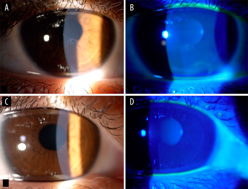 Figure 1. From left to right: (A) Right eye baseline examination: central epithelial pseudodendritic lesion, which (B) stained with fluorescein. (C) Left eye baseline examination: central epithelial pseudodentritic lesion which (D) faintly stained with fluorescein.
 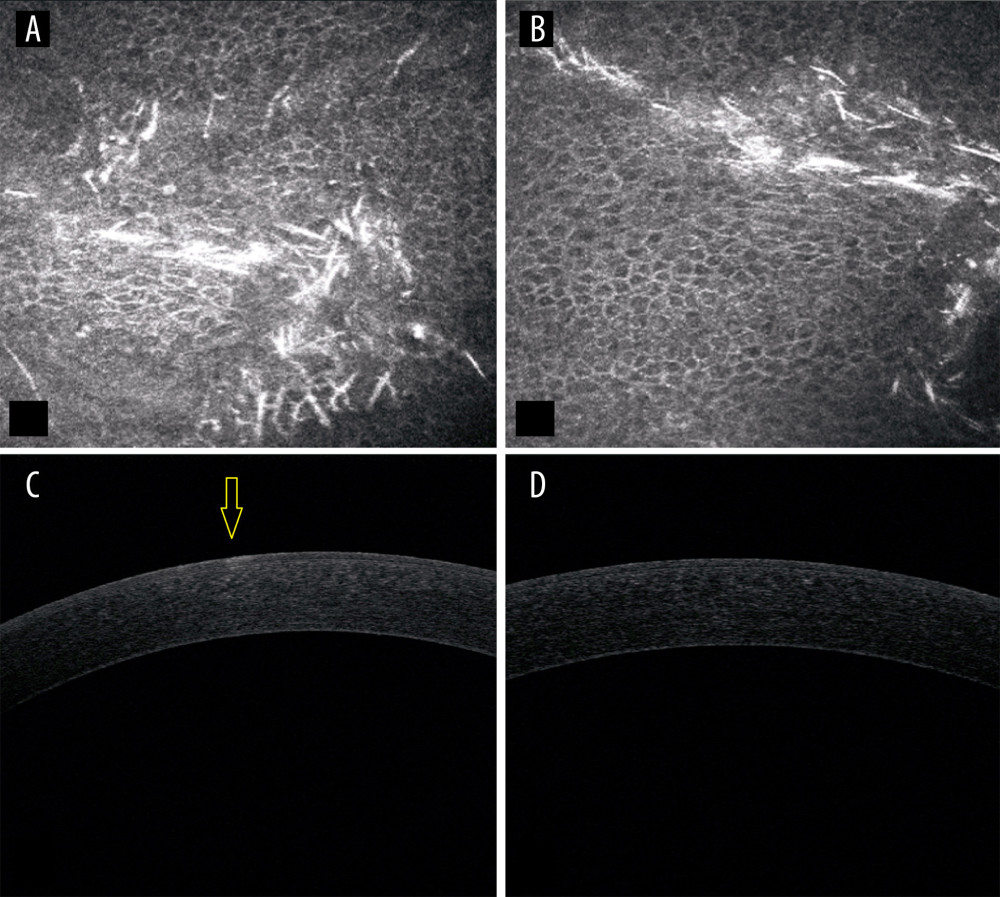 Figure 2. From left to right: (A) Right eye in vivo confocal microscopy (IVCM) baseline epithelial lesion. (B) Left eye IVCM baseline epithelial lesion (C) Right eye baseline anterior segment optical coherence tomography (AS-OCT) showing the lesion. (D) Left eye AS-OCT baseline without evidence of the lesion.
 References
1. Schiff M, Broue P, Chabrol BFrench-Belgian Study Group for HT-1, Heterogeneity of follow-up procedures in French and Belgian patients with treated hereditary tyrosinemia type 1: Results of a questionnaire and proposed guidelines: J Inherit Metab Dis, 2012; 35(5); 823-29
2. Chinsky JM, Singh R, Ficicioglu C, Diagnosis and treatment of tyrosinemia type I: A US and Canadian consensus group review and recommendations: Genet Med, 2017; 19(12); gim.2017.101
3. Bayzaei Z, Dehghani SM, Geramizadeh B, Tyrosinemia Type II Oct 24, 2024, Seattle (WA), University of Washington, Seattle
4. Barroso F, Correia J, Bandeira A, Tyrosinemia type III: A case report of siblings and literature review: Rev Paul Pediatr, 2020; 38; e2018158
5. Schauwvlieghe PP, Jaeken J, Kestelyn P, Claerhout I, Confocal microscopy of corneal crystals in a patient with hereditary tyrosinemia type I, treated with NTBC: Cornea, 2013; 32(1); 91-94
6. Kocabeyoglu S, Mocan MC, Irkec M, In vivo confocal microscopic features of corneal pseudodendritic lesions in tyrosinemia type II: Cornea, 2014; 33(10); 1106-8
7. Larrañaga Cores M, Domínguez García L, Estrada Vasquez N, Corneal crystals in patients with tyrosinemia types I and II: Cornea, 2024; 43(3); e4-e5
8. Kymionis GD, Kankariya VP, Kontadakis GA, Ziakas NG, Isolated corneal pseudodendrites as the initial manifestation of tyrosinemia type II in monozygotic twins: J Pediatr Ophthalmol Strabismus, 2012; 49; e33-36
9. Miranda BA, Rocha ACH, Arantes RR, Bilateral recurrent pseudodendritic keratopathy as the initial manifestation of tyrosinemia type II: Ophthalmic Genet, 2022; 43(2); 282-84
10. Gulmez Sevim D, Gumus K, Cavanagh HD, Corneal pseudodendritic lesions masquerading as herpetic keratitis in a patient with tyrosinemia type I: Eye Contact Lens, 2017; 43(3); e7-e9
Figures
Figure 1. From left to right: (A) Right eye baseline examination: central epithelial pseudodendritic lesion, which (B) stained with fluorescein. (C) Left eye baseline examination: central epithelial pseudodentritic lesion which (D) faintly stained with fluorescein.Figure 2. From left to right: (A) Right eye in vivo confocal microscopy (IVCM) baseline epithelial lesion. (B) Left eye IVCM baseline epithelial lesion (C) Right eye baseline anterior segment optical coherence tomography (AS-OCT) showing the lesion. (D) Left eye AS-OCT baseline without evidence of the lesion. In Press
Case report 
Am J Case Rep In Press; DOI: 10.12659/AJCR.953173
Case report 
Am J Case Rep In Press; DOI: 10.12659/AJCR.953192
Case report 
Am J Case Rep In Press; DOI: 10.12659/AJCR.952818
Case report
Am J Case Rep In Press; DOI: 10.12659/AJCR.953608
Most Viewed Current Articles
07 Dec 2021 : Case report
22,364,578
DOI :10.12659/AJCR.934347
Am J Case Rep 2021; 22:e934347
06 Dec 2021 : Case report  174,245
DOI :10.12659/AJCR.934406
Am J Case Rep 2021; 22:e934406
21 Jun 2024 : Case report
119,744
DOI :10.12659/AJCR.944371
Am J Case Rep 2024; 25:e944371
07 Mar 2024 : Case report
64,648
DOI :10.12659/AJCR.943133
Am J Case Rep 2024; 25:e943133