


27 August 2026: Articles 
Management Challenges of Interfering Cardiopulmonary Resuscitation-Induced Consciousness in the Prehospital Setting: A Case Report
Unusual clinical course, Challenging differential diagnosis, Management of emergency care
Nicholas A. Wright ABEF 1*, John B. Patterson AEF 2, William S. Cox AEF 3
DOI: 10.12659/AJCR.952977
Am J Case Rep 2026; 27:e952977


Abstract
BACKGROUND: Cardiopulmonary resuscitation-induced consciousness (CPRIC) is a rare phenomenon in which patients in cardiac arrest demonstrate signs of consciousness while undergoing cardiopulmonary resuscitation (CPR). Only a few cases of this phenomenon are reported in the literature. Previous studies estimate that CPRIC occurs in 0.23-0.9% of all cardiac arrests, with prevalence increasing over recent years. It has been proposed that the etiology of this phenomenon is the result of preserved cerebral perfusion during high-quality CPR.
CASE REPORT: A 52-year-old woman presented with witnessed out-of-hospital cardiac arrest after a bout of chest pain. During CPR, resuscitation was stopped multiple times due to signs of consciousness despite the patient being in ventricular fibrillation. Throughout the resuscitation attempt, the patient grunted, yelled, demonstrated purposeful movements, and opened her eyes. These actions resulted in considerable pauses in chest compressions, delays in routine advanced cardiac life support interventions, and psychological distress to the emergency medical services clinicians. After multiple defibrillations, the patient was successfully resuscitated and became oriented enough to answer questions and hold meaningful discussion. She was transported directly to the cardiac catheterization lab, where she underwent percutaneous coronary intervention to correct a 100% occlusive in-stent thrombosis of the mid-left anterior descending artery.
CONCLUSIONS: This case describes challenges health care professionals might face when CPRIC occurs. While sedation was considered, widespread protocols for sedation in CPRIC patients do not exist. More research is needed to determine the prevalence of CPRIC in the United States, as well as best practices for management of this rare phenomenon, for the safety of both patient and health care professionals.
Keywords: Cardiopulmonary Resuscitation, Case Reports, Consciousness, Emergency Medical Services, percutaneous coronary intervention, Ventricular Fibrillation
Introduction
Cardiac arrest is typically thought to manifest as a pulseless, apneic patient who is unresponsive. While this is true for the vast majority of patients, the prevalence of a phenomenon known as cardiopulmonary resuscitation-induced consciousness (CPRIC) is believed to have increased in recent years [1]. The phenomenon has been researched so little that, until recently, there has not even been a universally accepted definition of CPRIC, much less a consensus on how to manage it. In 2023, an international group of experts defined CPRIC as “a demonstration of consciousness while undergoing cardiopulmonary resuscitation with no measurable spontaneous cardiac output displayed. CPRIC can further be subgrouped into interfering and non-interfering CPRIC” [2]. This definition allows for identification of the phenomenon and lays the foundation for future research and management strategies.
Very few data are available regarding the etiology of the phenomenon or the reason for increased prevalence, but it has been speculated that increased cerebral blood flow during resuscitation may be the root cause [1]. Improved reporting of the phenomenon might play a role in the perceived increase in prevalence as well, though there is no way of knowing the true impact of this factor. Clinically, it would make sense that increased emphasis on high-quality cardiopulmonary resuscitation (CPR) in recent years, along with widespread implementation of mechanical CPR devices, might contribute to improved cerebral blood flow in cardiac arrest [3,4]. When paired with evidence that CPRIC patients have substantially higher rates of return of spontaneous circulation (ROSC) and 30-day survival, this explanation certainly seems to make physiologic sense [5].
Despite an absence of data regarding the prevalence of CPRIC in the United States, it is estimated that between 0.23% to 0.9% of all cardiac arrests globally present with signs of CPRIC [6]. When one considers that over 350 000 out-of-hospital cardiac arrests occur in the United States annually, prevalence should fall between 805 and 3150 cases per year if these estimations remain consistent. Despite these numbers, there are only a few case reports of the phenomenon in the literature [1,7–10]. The present case report describes a patient who presented as an out-of-hospital cardiac arrest and exhibited signs of interfering CPRIC. The report also describes how emergency services personnel responded, and discusses how improved awareness is crucial for appropriate management of CPRIC.
Case Report
Christiansburg Emergency Medical Services (CEMS) was dispatched to a 52-year-old woman in cardiac arrest. CEMS responded with an Advanced Life Support (ALS) ambulance and ALS response vehicle. New River Valley Emergency Communications Regional Authority (NRV 911) advised the responding crew that the patient was originally being driven to the emergency medical services (EMS) station by her significant other with the complaint of chest pain, but during the drive she became unresponsive. Telephone-assisted CPR instructions were provided through NRV 911 to the significant other. During the 4-minute response, NRV 911 updated the responding units that the patient was “awake and talking” and advised the significant other to stop CPR.
Upon arrival, the EMS crew found the patient supine on the ground, unresponsive, pulseless, and apneic, with the significant other actively performing chest compressions. No automated external defibrillator was available prior to EMS arrival. Due to environmental conditions that limited the ability to provide effective resuscitation outside, the patient was moved immediately into the ambulance and CPR was resumed without delay.
The patient’s medical history was significant for hypertension, hyperlipidemia, and an ST-elevation myocardial infarction that required balloon angioplasty and stenting of the mid-left anterior descending (LAD) artery. Medications documented by EMS included sublingual nitroglycerin, clopidogrel, and aspirin, though medication adherence was unclear. The patient also had a history of recreational cocaine use.
Shortly after CPR was resumed in the ambulance, the patient began moving her arms in a purposeful manner. Chest compressions were briefly paused to reassess for a pulse; however, the patient immediately became unresponsive when compressions stopped, and no pulse was identified. Compressions were immediately resumed, defibrillation pads were applied in the anterolateral position, the monitor was precharged to 200 J, and a rhythm check revealed ventricular fibrillation, prompting immediate defibrillation (Figure 1).
Chest compressions were resumed immediately while a simultaneous attempt was made to place a supraglottic airway. During compressions, the patient grunted, opened her eyes intermittently, and purposefully grabbed the compressor’s arms. Significant trismus prevented supraglottic airway placement. A nasopharyngeal airway was inserted to facilitate bag-valve-mask ventilations, which again elicited purposeful movement and eye opening. These findings prompted repeated reassessment for pulse and rhythm because the patient’s level of apparent awareness was inconsistent with the crew’s prior experiences managing cardiac arrests. Each time compressions were paused, however, the patient went completely limp, became unresponsive, and rhythm checks continued to demonstrate ventricular fibrillation. Defibrillation was delivered at appropriate intervals through a total of 6 shocks, although multiple brief pauses in compressions occurred due to the patient’s presentation and direct interference with resuscitative efforts, as well as the crew’s disbelief that the patient was in cardiac arrest, leading to the desire to confirm the underlying rhythm.
Vascular access was initially attempted intravenously due to concerns of potential intraosseous device dislodgment secondary to the patient flailing her limbs around during active resuscitation. Intraosseous access was deferred briefly while peripheral access was attempted. After a single unsuccessful intravenous attempt, intraosseous access was established in the left proximal tibia following the second defibrillation (Figure 1). The crew also cited concerns for patient discomfort as a reason for delaying intraosseous access, though this was not the primary factor influencing this decision.
After the third defibrillation (Figure 1), 4-lead electrocardiogram (ECG) monitoring was added as an additional means of confirming ventricular fibrillation. Ventricular fibrillation was confirmed in leads I, II, and III. Following the fourth defibrillation (Figure 1), a mechanical CPR device was applied and the patient’s arms were secured using the device straps to reduce interference with compressions. Despite this, the patient continued to exhibit purposeful movements, to the point of freeing her hands and attempting to pull the piston off her chest. She also opened her eyes and vocalized several obscenities with the device in place. At one point, the patient was able to push herself up on the stretcher enough to displace the device inferiorly, resulting in several compressions being delivered over the right costal margin before the device could be repositioned. Subsequent hospital imaging revealed multiple right-sided rib fractures, likely attributable to this event.
The receiving emergency department was contacted between the third and fourth defibrillations, and the attending physician was consulted regarding the patient’s awareness during CPR. The physician recommended the continuation of standard advanced cardiac life support measures. A subsequent call was placed to the same physician due to the severity of the patient’s agitation preventing effective resuscitation, at which time 2.5 mg of intraosseous midazolam was recommended. ROSC was achieved before this medication was administered.
Between the fifth and sixth defibrillations (Figure 1), the crew retrieved a second cardiac monitor from the ALS response vehicle and repeated a rhythm assessment to exclude monitor malfunction or artifact. The second monitor revealed identical findings to the first, confirming persistent ventricular fibrillation. This process contributed to a delay between the fifth and sixth shocks.
Shortly after the sixth defibrillation, the patient pushed herself upright in the mechanical CPR device and expressed significant distress, stating, “you’re all trying to kill me” and “you’re kidnapping me.” At this point, ROSC at been achieved. Vitals and a 12-lead ECG were obtained prior to transport. The patient was normotensive, tachycardic, and mildly hypoxic, with a Glascow Coma Score of 13 (E4, V4, M5). Hypoxia improved with 2 liters per minute of oxygen via nasal cannula. The 12-lead ECG was limited by motion artifact but demonstrated sinus tachycardia with significant septal and anterolateral ST-segment elevation (Figure 2). Based on these findings, the EMS crew immediately activated the cardiac catheterization lab (CCL) utilizing Pulsara, a smartphone application and “telehealth communication platform” that allows EMS personnel to directly activate and communicate with receiving CCL staff [11]. A repeat ECG was obtained during transport (Figure 3).
During transport, the patient remained agitated and repeatedly complained of dyspnea. On arrival at the receiving facility, the patient was taken directly to the CCL on the EMS stretcher.
The patient was intubated prior to transfer to the CCL table. Coronary angiography demonstrated a 100% occlusive in-stent thrombosis of the mid-LAD artery. Balloon angioplasty successfully restored flow and the patient remained hemodynamically stable throughout the procedure. The patient’s hospital course was otherwise uncomplicated, and she was discharged neurologically intact.
Discussion
This case highlights an extremely unusual presentation of a cardiac arrest patient exhibiting obvious signs of awareness and combativeness during resuscitation attempts, consistent with the interfering subtype of CPRIC. This patient required multiple rounds of defibrillation for refractory ventricular fibrillation and underwent at least 20 minutes of CPR with at least partial recall of the events. She was transported directly to the CCL for emergent percutaneous coronary intervention for an in-stent thrombosis of the LAD.
Many of the challenges faced by the EMS crew in this case have been reported in other cases of CPRIC, including purposeful movements of the extremities, grabbing of clinicians, vocalization, eye opening, and combativeness [1,7–10]. In this case, these behaviors complicated resuscitation efforts by prompting repeated pulse and rhythm reassessments, delaying vascular access, causing omission of advanced cardiac life support medication administration, and making airway management more difficult. The patient’s trismus prevented placement of an advanced airway and limited the ability to monitor end-tidal carbon dioxide, which has become an important metric during cardiac arrest management. A nasal cannula capable of monitoring end-tidal carbon dioxide was later applied, though no meaningful values were obtained until after ROSC.
Several interruptions in compressions occurred during the resuscitation; however, these pauses were generally brief and related to rhythm assessment or management of patient interference with ongoing CPR. Although it is not possible to determine the precise impact of these interruptions on CPR quality, the patient ultimately survived neurologically intact. This case underscores the importance of maintaining effective basic life support measures even in the setting of CPRIC. It is also possible that greater clinician familiarity with CPRIC may reduce unnecessary interruptions in future cases by increasing confidence that awareness and purposeful movement can occur despite ongoing cardiac arrest.
Point-of-care ultrasound may represent a useful adjunct in similar cases. One of the major challenges in this case was determining whether the patient remained in cardiac arrest despite repeated signs of awareness. Ultrasound could potentially have been used during an early rhythm check to assess for cardiac standstill or the absence of pulse-generating activity. Although ultrasound was available to the EMS crew, it was not utilized.
In addition to the barriers to patient care encountered during this case, there is also concern for physical injury and psychological stress among healthcare professionals that encounter CPRIC. In this case, one crew member sustained multiple abrasions and a hematoma to the forearm while performing compressions. The potential psychological effects of CPRIC on both clinicians and patients remain poorly studied and warrant further investigation [12].
Although the prevalence of CPRIC appears to be increasing, there have yet to be accepted guidelines or treatment algorithms developed for the care of these patients by major professional organizations. Likewise, little is known about the long-term psychological effects of CPRIC on patients. Patient and clinician safety should be the highest priority when developing these guidelines and future guidelines should prioritize both patient and clinician safety while minimizing interruptions in resuscitation. Chemical sedation may represent one strategy to reduce distress, purposeful movement, and interference with CPR, although evidence regarding its impact on outcomes remains limited. Several pharmacologic agents, including benzodiazepines, fentanyl, propofol, and ketamine, have been described in prior reports of CPRIC [2,6,13]. In some cases, including in this one, administration of both a sedative and a paralytic, such as succinylcholine or rocuronium, may warrant consideration when trismus interferes with airway management.
It is also important to consider that the use of sedation during CPRIC presents an ethical dilemma: clinicians may feel conflicted about sedating a patient who appears conscious and is “fighting for their life,” yet uncontrolled movement – let alone active resistance – interferes with almost every aspect of high-quality CPR. It is the authors’ opinion that sedation may not only be justified to reduce patient distress, but to also facilitate the delivery of high-quality resuscitation.
When considering sedation, one must consider the potential for hemodynamic compromise and how this may impact the patient both before and after ROSC. Ketamine may be particularly attractive in this setting because it can be administered intravenously, intraosseously, or intramuscularly, provides analgesia, and generally preserves hemodynamic stability even at sedating doses (1–2 mg/kg intravenous/intraosseous; 4–5 mg/kg intramuscular) [14–17]. Midazolam may be a reasonable alternative option, as it can also be administered through multiple routes and is generally hemodynamically tolerated when used alone, but it does not provide any analgesia [18,19]. The addition of an opioid such as fentanyl may improve analgesia, though the synergistic effects of these medications may increase the risk of hemodynamic compromise.
Delivering these medications during low-flow states (ie, CPR), particularly via tibial intraosseous access, also poses unique pharmacokinetic challenges. Delayed or unpredictable drug delivery can occur, as effective delivery relies on adequate perfusion for distribution to the sites of action, and this issue can complicate dose titration during CPR [20]. Inadequate circulation may also delay central drug delivery, which increases the risk of re-dosing these medications prior to the initial dose taking effect. This could theoretically present the potential for oversedation after ROSC.
While ketamine and midazolam have been utilized successfully in other cases, further investigation is needed to determine optimal medication selection, dosing, and safety in patients with CPRIC.
Conclusions
CPRIC is a challenging and increasingly recognized phenomenon for which no universal guidelines for appropriate management or sedation currently exist [2,5]. This case illustrates one possible presentation of CPRIC, as well as the barriers to care encountered by clinicians. Increased awareness of CPRIC among healthcare professionals may help clinicians to better recognize the phenomenon and respond more effectively when it occurs. Further research is needed to clarify the prevalence of CPRIC in the United States, identify optimal sedation strategies and dosing, develop evidence-based management guidelines, and better understand the long-term psychological effects on both survivors and healthcare professionals.
Figures
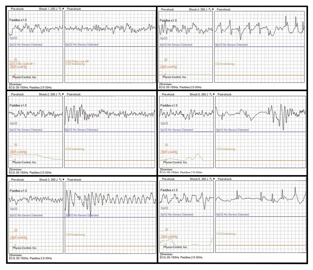 Figure 1. The initial defibrillation was delivered at 200 joules for ventricular fibrillation (top left). The second defibrillation was delivered at 300 joules for ventricular fibrillation (middle left). The third defibrillation was delivered at 360 joules for ventricular fibrillation, and the rhythm appears to have converted into ventricular tachycardia post-defibrillation (lower left). The fourth defibrillation was delivered at 360 joules for ventricular fibrillation, and the rhythm appears to have converted into an organized rhythm for a brief period of time post-shock (top right). The fifth defibrillation was delivered at 360 joules for ventricular fibrillation, and the rhythm appears to have once again converted into an organized rhythm for a brief period post-shock (middle right). The sixth defibrillation was delivered at 360 joules for ventricular fibrillation, and ROSC was obtained post-shock (lower right). ROSC, return of spontaneous circulation.
 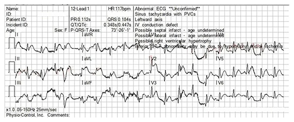 Figure 2. First 12-lead ECG post-ROSC. The ECG was complicated by significant artifacts due to patient noncompliance. ST-segment elevation was significant enough that emergency medical services clinicians activated the CCL based on these findings. ECG, electrocardiogram; ROSC, return of spontaneous circulation; CCL, cardiac catheterization lab.
 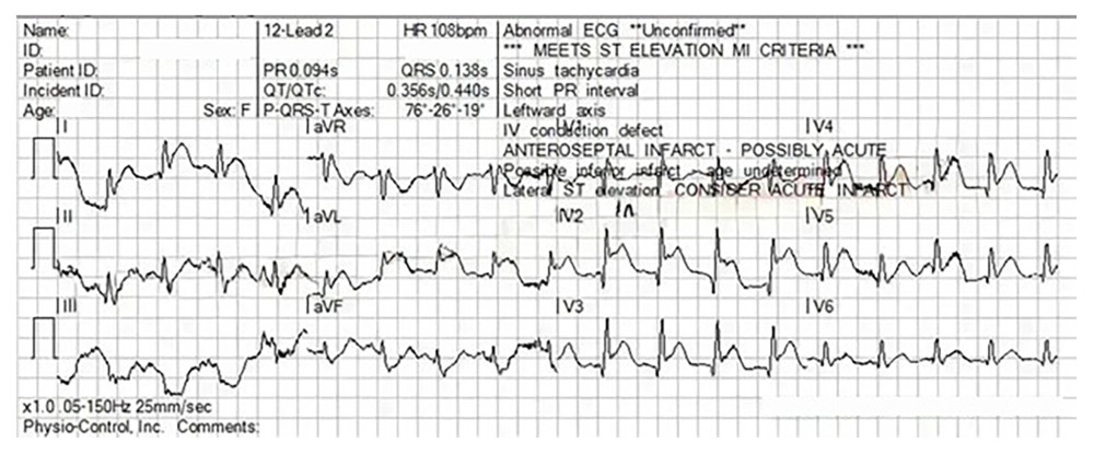 Figure 3. Second 12-lead ECG. There is a marked improvement in artifact levels with this ECG. This demonstrates clear ST-segment elevation in the anteroseptal leads with reciprocal depression. ECG, electrocardiogram.
 References
1. Olaussen A, Shepherd M, Nehme Z, Return of consciousness during ongoing cardiopulmonary resuscitation: A systematic review: Resuscitation, 2015; 86; 44-48
2. Howard J, Grusd E, Rice D, Development of an international prehospital CPR-induced consciousness guideline: A Delphi study: Paramedicine, 2024; 21(2); 66-78
3. Rubertsson S, Karlsten R, Increased cortical cerebral blood flow with LUCAS: A new device for mechanical chest compressions compared to standard external compressions during experimental cardiopulmonary resuscitation: Resuscitation, 2005; 65(3); 357-63
4. Obermaier M, Zimmermann JB, Popp E, Automated mechanical cardiopulmonary resuscitation devices versus manual chest compressions in the treatment of cardiac arrest: Protocol of a systematic review and meta-analysis comparing machine to human: BMJ Open, 2021; 11(2); e042062
5. Doan TN, Adams L, Schultz BV, Insights into the epidemiology of cardiopulmonary resuscitation-induced consciousness in out-of-hospital cardiac arrest: Emerg Med Australas, 2020; 32(5); 769-76
6. West RL, Otto Q, Drennan IR, CPR-related cognitive activity, consciousness, awareness and recall, and its management: A scoping review: Resuscitation Plus, 2022; 10; 100241
7. Pound J, Verbeek PR, Cheskes S, CPR-induced consciousness during out-of-hospital cardiac arrest: A case report on an emerging phenomenon: Prehosp Emerg Care, 2016; 21(2); 252-56
8. Rice D, Nudell N, Habrat D, CPR-induced consciousness: Sedation protocols for this special population: Br Paramed J, 2016; 1(2); 24-30
9. Pinto J, Almeida P, Ribeiro F, Cardiopulmonary resuscitation-induced consciousness: A case report in an elderly patient: European J Case Rep Internal Med, 2020; 7(2); 001409
10. Al Atbi A, Al Mandhari A, Al Reesi A, Cardiopulmonary resuscitation-induced consciousness: A case report: Oman Med J, 2022; 37(2); e351
11. Pulsara: About Pulsara [Internet], Bozeman (MT), Pulsara [cited 2024 Feb 1]. Available from: https://www.pulsara.com/about
12. Parnia S, Shirazi T, Patel J, AWAreness during REsuscitation II: A multicenter study of consciousness and awareness in cardiac arrest: Resuscitation, 2023; 191; 109903
13. Pourmand A, Hill B, Yamane D, Approach to cardiopulmonary resuscitation-induced consciousness: An emergency medicine perspective: Am J Emerg Med, 2019; 37(4); 751-56
14. Scheppke K, Braghiroli J, Chalaby M, Prehospital use of intramuscular ketamine for sedation of violent and agitated patients: Western J Emerg Med, 2014; 15(7); 736-41
15. Linder LM, Ross CA, Weant KA, Ketamine for the acute management of excited delirium and agitation in the prehospital setting: Pharmacotherapy, 2018; 38(1); 139-51
16. Banik S, Madavi S, Exploring the role of ketamine sedation in critically ill patients: A comprehensive review: Cureus, 2024; 16(7); e65836
17. Strayer RJ, Nelson LS, Adverse events associated with ketamine for procedural sedation in adults: Am J Emerg Med, 2008; 26(9); 985-1028
18. Frölich MA, Arabshahi A, Katholi C, Hemodynamic characteristics of midazolam, propofol, and dexmedetomidine in healthy volunteers: J Clin Anesthesia, 2011; 23(3); 218-23
19. Cernaianu AC, DelRossi AJ, Flum DR, Lorazepam and midazolam in the intensive care unit: A randomized, prospective, multicenter study of hemodynamics, oxygen transport, efficacy, and cost: Crit Care Med, 1996; 24(2); 222-28
20. Hoskins SL, do Nascimento P, Lima RM, Pharmacokinetics of intraosseous and central venous drug delivery during cardiopulmonary resuscitation: Resuscitation, 2012; 83(1); 107-12
Figures
Figure 1. The initial defibrillation was delivered at 200 joules for ventricular fibrillation (top left). The second defibrillation was delivered at 300 joules for ventricular fibrillation (middle left). The third defibrillation was delivered at 360 joules for ventricular fibrillation, and the rhythm appears to have converted into ventricular tachycardia post-defibrillation (lower left). The fourth defibrillation was delivered at 360 joules for ventricular fibrillation, and the rhythm appears to have converted into an organized rhythm for a brief period of time post-shock (top right). The fifth defibrillation was delivered at 360 joules for ventricular fibrillation, and the rhythm appears to have once again converted into an organized rhythm for a brief period post-shock (middle right). The sixth defibrillation was delivered at 360 joules for ventricular fibrillation, and ROSC was obtained post-shock (lower right). ROSC, return of spontaneous circulation.Figure 2. First 12-lead ECG post-ROSC. The ECG was complicated by significant artifacts due to patient noncompliance. ST-segment elevation was significant enough that emergency medical services clinicians activated the CCL based on these findings. ECG, electrocardiogram; ROSC, return of spontaneous circulation; CCL, cardiac catheterization lab.Figure 3. Second 12-lead ECG. There is a marked improvement in artifact levels with this ECG. This demonstrates clear ST-segment elevation in the anteroseptal leads with reciprocal depression. ECG, electrocardiogram. In Press
Case report 
Am J Case Rep In Press; DOI: 10.12659/AJCR.953317
Case report 
Am J Case Rep In Press; DOI: 10.12659/AJCR.954643
Case report 
Am J Case Rep In Press; DOI: 10.12659/AJCR.954074
Case report
Am J Case Rep In Press; DOI: 10.12659/AJCR.953592
Most Viewed Current Articles
07 Dec 2021 : Case report
28,712,429
DOI :10.12659/AJCR.934347
Am J Case Rep 2021; 22:e934347
06 Dec 2021 : Case report  180,530
DOI :10.12659/AJCR.934406
Am J Case Rep 2021; 22:e934406
21 Jun 2024 : Case report
124,276
DOI :10.12659/AJCR.944371
Am J Case Rep 2024; 25:e944371
08 May 2026 : Case report
95,045
DOI :10.12659/AJCR.952158
Am J Case Rep 2026; 27:e952158