


15 September 2026: Articles 
A Case of Pneumocephalus on Valsalva Maneuver: A Case Report and Literature Review
Unusual clinical course
Bouchra Chergui EF 1, Julien Flament E 2, Nicolas Mulquin B 3, Céline Couvreur E 1*
DOI: 10.12659/AJCR.953059
Am J Case Rep 2026; 27:e953059


Abstract
BACKGROUND: Pneumocephalus is defined as the presence of air within the cranial cavity. Spontaneous onset is rare, with most cases arising secondary to head trauma or surgical procedures. The most common symptom is headache, a frequent concern in the emergency department, with a broad differential diagnosis. The diagnosis is based on a simple brain computed tomography (CT), while the etiological assessment involves magnetic resonance imaging (MRI) and consultation with otorhinolaryngologists. Most cases of pneumocephalus can be managed conservatively; however, some require urgent surgical intervention.
CASE REPORT: We report the case of a 45-year-old woman with no history of recent trauma or neurosurgical procedures who developed a sudden-onset occipital headache following a Valsalva maneuver. The pain worsened with head movement. Neurological and otorhinolaryngological examinations were unremarkable. Brain CT revealed a small, isolated pneumocephalus localized to the posterior fossa. No anatomical defect was identified on CT and MRI. The patient was managed conservatively. Her symptoms progressively resolved, and follow-up imaging demonstrated spontaneous resorption of the intracranial air. The favorable course suggests spontaneous closure of a potentially Valsalva-induced intracranial sinus cavity.
CONCLUSIONS: This case highlights the importance of neuroimaging in patients presenting with atypical headaches in the emergency department. Despite its rarity, pneumocephalus remains an underrecognized condition in emergency medicine. In addition to describing an unusual presentation, this report includes a review of previously published cases of Valsalva-induced spontaneous pneumocephalus and contributes a further example to the limited literature on this subject.
Keywords: Emergency Medicine, Headache, Pneumocephalus, Valsalva Maneuver
Introduction
Pneumocephalus, also known as pneumocranium, is defined as the presence of air or gas within the cranial cavity. It is an uncommon condition, most frequently associated with trauma, neurosurgical procedures, infections, or otorhinolaryngological interventions. Spontaneous pneumocephalus is relatively rare, representing approximately 0.6% of all cases, and remains a poorly understood phenomenon [1]. Clinical manifestations vary according to the volume of intracranial air, its location, and the rapidity of onset. Headaches are the most common presenting symptom; however, confusion, lethargy, focal neurological deficits, meningeal signs, dizziness, nausea, and vomiting can also occur [2].
We describe the case of a patient who presented to the emergency department with an atypical headache following a Valsalva maneuver, ultimately diagnosed as pneumocephalus. Given the rarity of spontaneous pneumocephalus and its often non-specific clinical presentation, reporting this case together with a review of previously published cases adds to the existing literature and may assist clinicians in recognizing this uncommon condition, especially when triggered by a Valsalva maneuver.
Case Report
A 45-year-old woman presented at our emergency department with persistent headaches following a Valsalva maneuver, which she had performed to relieve a sensation of ear fullness. The headache was constant, non-pulsatile, localized to the occipital region, and exacerbated by movement. She also reported photophobia but denied nausea or vomiting. Clinical examination, including a detailed neurological assessment, was unremarkable. The patient had a history of migraines with aura but described this headache as distinct from her usual migraine episodes. She had no other known past medical history and was not receiving any ongoing treatment.
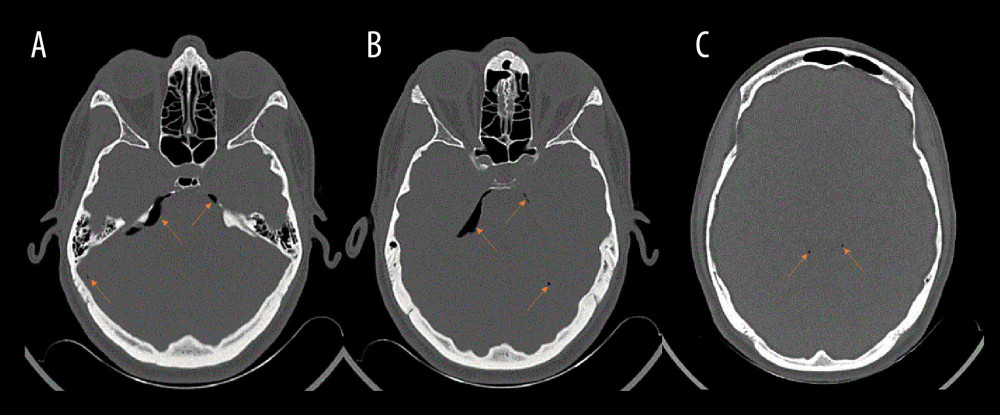
A non-contrast brain CT scan revealed an isolated pneumocephalus in the posterior fossa, with no identifiable cause (such as sign of trauma or sinus pathology) (Figure 1). Given the risk of intracranial infection, empirical antibiotic therapy with amoxicillin/clavulanic acid was initiated but subsequently switched to clindamycin following an allergic reaction. She was admitted to the neurology department for observation. Brain magnetic resonance imaging (MRI) performed 2 days later demonstrated residual pneumocephalus beneath the tentorium cerebelli and in the cerebellopontine angle cisterns, but no evidence of fistula or petrous apex abnormalities. Spinal MRI and anterior rhinoscopy were also unremarkable.
Following multidisciplinary discussion, the leading hypothesis was a transient opening of an intracranial sinus-like cavity induced by the Valsalva maneuver, possibly due to a pre-existing weakness in the cavity wall. The favorable clinical and radiological evolution suggested spontaneous closure. At a follow-up consultation 6 weeks later, she remained asymptomatic except for persistent right ear fullness, with no otolaryngological abnormalities detected. Repeat brain CT confirmed complete resolution of the pneumocephalus.
Discussion
The first cases of pneumocephalus were reported after head trauma or surgery. In 1884, Chiari described pneumocephalus post-mortem in a patient with ethmoiditis induced by a forced Valsalva maneuver [3]. Since 1970s, the advent of the CT imaging has considerably improved its diagnosis.
The literature distinguishes several subtypes of spontaneous pneumocephalus, including tension and non-tension pneumocephalus, each with specific clinical implications and management. Non-tension pneumocephalus is usually asymptomatic and resolves spontaneously, whereas tension pneumocephalus is a rare but severe neurosurgical emergency caused by intracranial air exerting pressure on the brain parenchyma [4]. Pneumocephalus is also classified anatomically as extradural, subdural, subarachnoidal, intraparenchymal, or intraventricular [5]. Based on the clinical and radiological findings in this case, the pneumocephalus observed was intradural, specifically located in the posterior fossa beneath the tentorium cerebelli and within the cerebellopontine angle cisterns. In the absence of trauma, surgical intervention, or identifiable sinus pathology, and given the spontaneous resolution, this case corresponds to spontaneous non-tension pneumocephalus. Although some case reports describe pneumocephalus as minor, moderate, or massive, there is no clear classification based on volume [6]. The clinical significance lies in the physiological impact of intracranial air, particularly in cases of tension pneumocephalus, where mass effect can result in neurological deterioration.
Two mechanisms have been proposed to explain spontaneous pneumocephalus [7]:
The underlying mechanism of pneumocephalus in our patient corresponds to the ball-valve mechanism.
In most cases, non-tension pneumocephalus is asymptomatic and resolves spontaneously. However, some patients have sudden, severe headache, sometimes accompanied by nausea and vomiting. Cerebrospinal fluid rhinorrhea can also occur. Patients can present with neurological signs such as confusion, altered consciousness, or, less often, focal neurological deficits mimicking stroke. Seizures are rare but reported [8]. Additional symptoms can include otalgia, tinnitus, imbalance, and a sensation of aural fullness. Meningeal irritation signs may be found on examination. A rare but pathognomonic sign is a splashing sound synchronous with head movement [1], a cerebrospinal splash-like noise detectable during head movements, which indicates the coexistence of intracranial air and fluid.
The diagnosis of pneumocephalus is primarily based on imaging, with non-contrast CT as the gold standard. CT precisely visualizes intracranial air and its location, detecting volumes as small as 0.5 mL [9]. This high sensitivity is due to the marked density difference between air and brain tissue, allowing even very small volumes of pneumocephalus to be rapidly identified [6]. CT is also essential for assessing severity, particularly in identifying tension pneumocephalus, a neurosurgical emergency. In addition, CT helps exclude other urgent causes of sudden headache, such as subarachnoid hemorrhage. In our case, CT revealed a non-tension pneumocephalus with air confined to the posterior fossa, without skull fracture or sinus pathology. MRI is useful for follow-up but is less sensitive for intracranial air detection [6]. However, MRI is valuable for identifying underlying structural abnormalities, such as meningeal fistulas or skull base malformations. Other investigations can be relevant for establishing the underlying etiology. Cerebrospinal fluid leaks can be assessed by measuring biomarkers, particularly in nasal secretions [10]. In cases of high suspicion, when laboratory tests come back negative, MRI (or CT) cisternography with an intrathecal injection of gadolinium can be useful [11]. A detailed CT scan, such as one focused on the temporal bone, can be helpful in locating a bone defect, particularly in preparation for surgical treatment [10]. Multidisciplinary collaboration with otorhinolaryngologists is essential. Endoscopic examination and otogenic evaluation allow exclusion of a sinus lesion and identification of an otogenic source of pneumocephalus, respectively, particularly in cases of mastoid hyperpneumatization [12]. In our case, these otorhinolaryngology evaluations were performed.
Uncomplicated pneumocephalus is generally managed conservatively. Strict bed rest in the supine position is recommended to minimize intracranial pressure. High-flow oxygen therapy, delivered by a non-rebreather mask or other systems, promotes intracranial air resorption through a diffusion gradient mechanism, in accordance with Fick’s law [13]. This often results in early clinical and radiological improvement. In severe cases, hyperbaric oxygen therapy may accelerate intracranial air reduction and symptoms relief [14]. Surgery is rarely required in cases of benign pneumocephalus. The indications for surgical management are tension pneumocephalus, failure of conservative treatment, and presence of a significant bony defect. Endoscopic endonasal surgery is the gold standard for treating anterior skull base leaks, with reported success rates exceeding 90% [15]. This minimally invasive approach relies on precise localization and exposure of the bony defect, followed by multilayer reconstruction using autologous grafts. For larger defects, vascularized pedicled flaps improve repair durability and reduce recurrence risk. For temporal bone defects—particularly involving the tegmen tympani or squamous portion—a middle cranial fossa approach may be indicated. This involves a craniotomy with extradural exposure to identify bony and dural defects, followed by multilayer dural repair and reconstruction. In emergency situations, especially with tension intraventricular pneumocephalus, ventriculostomy may be required for rapid intracranial decompression prior to definitive surgical repair [16].
In most cases, pneumocephalus resolves spontaneously. Air resorption typically occurs within a few days to 2 to 3 weeks, especially when the air volume is small and no fistula persists. There are no clear recommendations regarding the duration of follow-up in cases managed conservatively. However, the available literature consistently supports close clinical and radiological monitoring. Spontaneous resolution occurs in approximately 80% to 90% of reported cases without complications or severity factors. Prognosis depends on the underlying etiology. Spontaneous or infectious forms may have a poorer outcome, particularly if diagnosis and treatment is delayed. The most serious complication is tension pneumocephalus (< 5% of cases), which can lead to rapid neurological deterioration, requiring urgent surgical intervention [17]. Other complications include infections (meningitis or brain abscesses), reported in 5% to 10% of cases, especially in post-traumatic or postoperative pneumocephalus associated with a dural breach [18]. This explains why our patient initially received antibiotic prophylaxis.
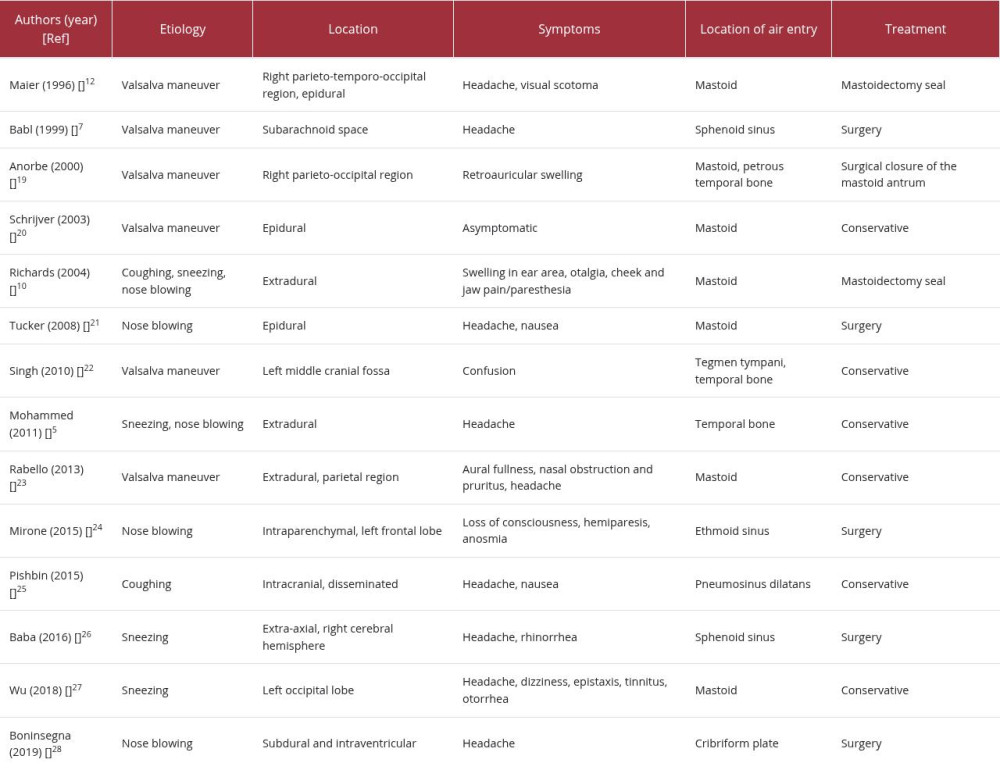
Spontaneous intracerebral pneumocephalus of otogenic origin is a rare condition, with to our knowledge only 14 cases reported in the medical literature. We conducted a literature search to identify cases of spontaneous pneumocephalus induced by Valsalva maneuver. The search was performed in PubMed up to October 2024. We used the keywords “spontaneous pneumocephalus”, “Valsalva maneuver”, and “nontraumatic pneumocephalus”. References cited within the selected papers were also reviewed to identify additional relevant reports. This cases of spontaneous intracerebral pneumocephalus of otogenic origin are listed in Table 1.
Conclusions
Pneumocephalus often remains undetected due to its non-specific clinical presentation. The advent of CT has significantly improved diagnosis accuracy, allowing rapid detection and precise localization of intracranial air. This case of pneumocephalus following a Valsalva maneuver illustrates the importance of prompt recognition and diagnosis to ensure appropriate management. Our case adds to the limited literature on spontaneous pneumocephalus and highlights the potential for spontaneous resolution in selected cases. It also underscores the importance of multidisciplinary management and close clinical and radiological follow-up. However, within the limited literature on this topic, the mechanisms underlying the development of spontaneous pneumocephalus remain unclear and warrant further investigation in future studies.
References
1. Markham JW, The clinical features of pneumocephalus based upon a survey of 284 cases with report of 11 additional cases: Acta Neurochir (Wien), 1967; 16; 1-78
2. Wankhade BS, Beniamein MMK, Alrais ZF, What should an intensivist know about pneumocephalus and tension pneumocephalus?: Acute Crit Care, 2023; 38(2); 244-48
3. Chiari HA case of accumulation of air in the ventricles of human brain: Zeitschrift für Heilkunde, 1884; 5; 383-90 [in German]
4. Sweni S, Senthilkumaran S, Balamurugan N, Tension pneumocephalus: A case report with review of literature: Emerg Radiol, 2013; 20; 573-78
5. Mohammed el R, Profant M, Spontaneous otogenic pneumocephalus: Acta Otolaryngol, 2011; 131(6); 670-74
6. Schirmer CM, Heilman CB, Bhardwaj A, Pneumocephalus: Case illustrations and review: Neurocrit Care, 2010; 13; 152-58
7. Babl FE, Arnett AM, Barnett P, Sherwood M, Atraumatic pneumocephalus: A case report and review of the literature: Pediatr Emerg Care, 1999; 15(2); 106-9
8. Rattu MA, Aziz M, Powell K, Pneumocephalus – Epidural injection nightmare: Research Posters, 2024
9. Vacca VM, Pneumocephalus assessment and management: Nursing Crit Care, 2017; 12(4); 24-29
10. Richards SD, Saeed SR, Laitt R, Ramsden RT, Hypercellularity of the mastoid as a cause of spontaneous pneumocephalus: J Laryngol Otol, 2004; 118(6); 474-76
11. Eljazzar R, Loewenstern J, Dai JB, Detection of cerebrospinal fluid leaks: Is there a radiologic standard of care? A systematic review: World Neurosurg, 2019; 127; 307-15
12. Maier W, Fradis M, Scheremet R, Spontaneous otogenic pneumocephalus: Ann Otol Rhinol Laryngol, 1996; 105(4); 300-2
13. Kim BJH, Lee JH, Moon KS, Use of oxygen therapy for pneumocephalus: A systematic review: Neurosurg Rev, 2024; 47; 1-6
14. Paiva W, de Andrade AF, Amorim RL, Effects of hyperbaric oxygenation therapy on symptomatic pneumocephalus: Ther Clin Risk Manag, 2014; 10; 769-73
15. Gâta A, Toader C, Trombitaş VE, Endoscopic skull base repair strategy for CSF leaks associated with pneumocephalus: J Clin Med, 2020; 10(1); 46
16. Piccirilli M, Pescatori L, Grasso G, Timing of surgical intervention in anterior frontobasal fractures with pneumocephalus and cerebrospinal fluid leaks: A retrospective analysis on a series of 314 patients and proposal of a novel classification and management algorithm: World Neurosurgery, 2025; 201; 124353
17. Gorissen Z, Hakvoort K, Van Den Boogaart M, Pneumocephalus: A rare and life-threatening, but reversible, complication after penetrating lumbar injury: Acta Neurochir (Wien), 2019; 161; 361-65
18. Śmilowska K, Sznajder-Stacha K, Kocyłowski D, Pneumocephalus as a rare complication: A systematic review plus clinical vignette: Pol J Neurol Neurosurg, 2021; 55(3); 253-68
19. Anorbe E, Aisa P, Saenz de Ormijana J, Spontaneous pneumatocele and pneumocephalus associated with mastoid hyperpneumatization: Eur J Radiol, 2000; 36(1); 45-48
20. Schrijver HM, Berendse HW, Pneumocephalus by Valsalva’s maneuver: Neurology, 2003; 60(2); 345-46
21. Tucker A, Miyake H, Tsuji M, Spontaneous epidural pneumocephalus: Neurol Med Chir (Tokyo), 2008; 48(10); 474-78
22. Singh A, Alvarez J, Spontaneous otogenic intracerebral pneumocephalus: West J Emerg Med, 2010; 11(1); 107
23. Rabello FA, Massuda ET, de Oliveira JAA, Hyppolito MA, Otogenic spontaneous pneumocephalus: Case report: Braz J Otorhinolaryngol, 2013; 79(5); 643
24. Mirone G, Rotondo M, Scuotto A, Spontaneous intraparenchymal tension pneumoce-phalus triggered by compulsive forceful nose blowing: Am J Emerg Med, 2015; 33(9); 1320e1-3
25. Pishbin E, Jabbari M, Parsa A, Spontaneous nontraumatic pneumocephalus: A case report: Iran Red Crescent Med J, 2015; 17(7); e23920
26. Baba M, Tarar O, Syed A, A rare case of spontaneous pneumocephalus associated with nontraumatic cerebrospinal fluid leak: Case Rep Neurol Med, 2016; 2016; 1828461
27. Wu YH, Yeh IJ, Liu KT, Spontaneous pneumocephalus and subdural hemorrhage after sneezing: Am J Emerg Med, 2018; 36(10); 1928e1-3
28. Boninsegna E, Barillari M, De Rossi S, Non-traumatic pneumocephalus caused by increased depth of olfactory fossa: Br J Oral Maxillofac Surg, 2019; 57(6); 587-89
In Press
Case report 
Am J Case Rep In Press; DOI: 10.12659/AJCR.953543
Case report 
Am J Case Rep In Press; DOI: 10.12659/AJCR.954216
Case report 
Am J Case Rep In Press; DOI: 10.12659/AJCR.954932
Case report 
Am J Case Rep In Press; DOI: 10.12659/AJCR.954146
Most Viewed Current Articles
07 Dec 2021 : Case report
28,712,748
DOI :10.12659/AJCR.934347
Am J Case Rep 2021; 22:e934347
06 Dec 2021 : Case report  180,779
DOI :10.12659/AJCR.934406
Am J Case Rep 2021; 22:e934406
21 Jun 2024 : Case report
124,457
DOI :10.12659/AJCR.944371
Am J Case Rep 2024; 25:e944371
08 May 2026 : Case report
95,565
DOI :10.12659/AJCR.952158
Am J Case Rep 2026; 27:e952158