


21 August 2026: Articles 
Left Ventricular Summit Ventricular Tachycardia Identified by Electrocardiographic Pattern Recognition and Managed With Radiofrequency Ablation
Challenging differential diagnosis, Management of emergency care, Rare disease
Mariana Goes Moreira ABCDEF 1, Carina Abigail Hardy ABD 1, Leandro Menezes Alves da Costa ACD 1, Ana Carolina Menezes Borsoi ACD 1, Rodrigo Goldenstein Schainberg ACD 1, Anna Beatriz Gori Montes ACD 1, Rafael Amorim Belo Nunes ACD 1, Thiago Midlej Brito ACD 1, Daniel Castanho Genta Pereira ACD 1, Roger Pereira de Oliveira ACD 1, Thiago Luis Scudeler ABCDEF 1*
DOI: 10.12659/AJCR.953321
Am J Case Rep 2026; 27:e953321


Abstract
BACKGROUND: Left ventricular summit ventricular tachycardia (LVSVT) originates in the epicardium of the superior left ventricular wall, between the origins of the main coronary arteries, with characteristic findings on 12-lead electrocardiogram (ECG) that can guide ablation. This report describes a 66-year-old man with dizziness and palpitations diagnosed with LVSVT using ECG and managed with radiofrequency ablation.
CASE REPORT: A 66-year-old man presented with 24 hours of dizziness and palpitations. He was hemodynamically stable, and the initial ECG showed frequent premature ventricular complexes with left bundle branch block-like morphology, inferior axis, and early precordial transition. Continuous monitoring and 24-hour Holter recording documented a high ventricular ectopic burden (~65%), with episodes of sustained and nonsustained monomorphic ventricular tachycardia of identical morphology. Echocardiography showed preserved left ventricular systolic function, and coronary angiography and cardiac magnetic resonance imaging excluded obstructive coronary disease, myocardial fibrosis, and scar. Because symptoms and arrhythmia burden persisted, an electrophysiological study was performed on day 4. Isoproterenol infusion induced ventricular tachycardia, activation mapping localized the earliest ventricular activation to the left ventricular summit, and radiofrequency ablation was performed from the great cardiac vein and adjacent left ventricular outflow tract and left coronary cusp sites after coronary angiography confirmed a safe distance from the coronary arteries. Ventricular ectopy was immediately suppressed, and no arrhythmia was inducible after ablation. At 30-day follow-up, the patient remained asymptomatic without recurrent ventricular arrhythmia on Holter monitoring.
CONCLUSIONS: Systematic ECG interpretation can localize LVSVT and guide effective catheter ablation.
Keywords: Bundle-Branch Block, Electrocardiography, Electrophysiology, Tachycardia, Ventricular
Introduction
Ventricular arrhythmias arising from the left ventricular summit are an important subset of idiopathic outflow tract ventricular arrhythmias and are challenging because of the region’s epicardial anatomy, adjacency to the left main coronary bifurcation, and relationship with the coronary venous system [1,2]. The left ventricular summit is the most superior epicardial aspect of the left ventricle and is usually described as a triangular region bounded by the left anterior descending artery, the left circumflex artery, and the great cardiac vein or anterior interventricular vein [1,2]. The great cardiac vein divides the region into an inferior area that may be accessible to catheter ablation and a superior area that is often inaccessible because of coronary artery proximity and epicardial adipose tissue [2].
Left ventricular summit ventricular arrhythmias account for approximately 10% to 15% of idiopathic ventricular arrhythmias and may present as premature ventricular complexes (PVCs), nonsustained ventricular tachycardia (NSVT), or sustained ventricular tachycardia (SVT) [1,2]. Patients can present with palpitations, dizziness, presyncope, syncope, or incidentally detected frequent ectopy; a high ectopic burden can also contribute to left ventricular dysfunction in susceptible patients [1,3]. Diagnosis begins with a 12-lead electrocardiogram (ECG) and ambulatory rhythm monitoring and is refined by imaging to exclude ischemic or structural disease and by electrophysiological mapping when ablation is planned [1,4].
Electrocardiographic localization is clinically useful because left ventricular summit arrhythmias often show inferior-axis outflow tract patterns, early precordial transition, and limb-lead features suggesting an anterosuperior and frequently epicardial origin [1,4]. However, the electrocardiographic appearance may overlap with right ventricular outflow tract, aortic cusp, left ventricular outflow tract, and coronary venous system origins; therefore, electrocardiographic interpretation must be integrated with coronary imaging and 3-dimensional mapping [1,4–6].
Management depends on symptoms, arrhythmia burden, structural heart disease, and procedural risk. Medical therapy may be used, but catheter ablation is considered when symptoms or ectopy burden persist or when ventricular tachycardia is recurrent [1,3]. Ablation can require direct energy delivery from the great cardiac vein or anterior interventricular vein, an indirect approach from the left ventricular outflow tract, aortic cusps, or adjacent endocardial structures, or advanced strategies when conventional radiofrequency ablation is limited by coronary artery proximity [1–3,6]. A previously published case report described successful ablation of a left ventricular summit ventricular tachycardia (LVSVT) focus through the left atrial appendage, emphasizing that individualized access routes may be required for this region [5]. The present report describes the case of a 66-year-old man with dizziness and palpitations diagnosed with LVSVT using electrocardiography and managed with radiofrequency ablation.
Case Report
A 66-year-old man presented to the emergency department with a 24-hour history of dizziness and palpitations. He denied syncope, chest pain, or dyspnea. At presentation, he was hemodynamically stable, with blood pressure of 122/82 mm Hg and heart rate of 50 beats/min. Physical examination was unremarkable.
The patient’s medical history included hypertension, type 2 diabetes mellitus, and coronary artery disease treated with percutaneous coronary intervention of the left anterior descending artery 7 years earlier. His chronic medications were aspirin, rosuvastatin, metformin, and losartan. There was no clinically relevant history of tobacco, alcohol, or substance abuse documented in the medical record.
The initial 12-lead ECG showed sinus rhythm with frequent PVCs (Figure 1). The PVCs had left bundle branch block-like morphology, inferior axis, and early precordial transition. A subsequent ECG obtained in the intensive care unit showed episodes of NSVT with identical QRS morphology (Figure 2), indicating a shared arrhythmogenic focus. Limb-lead analysis showed higher R-wave amplitude in lead II than in lead III and absence of septal q waves, findings that further supported an anterosuperior and likely epicardial site of origin.
Transthoracic echocardiography showed preserved left ventricular ejection fraction of 57%, with no regional wall motion abnormality or clinically significant valvular disease. Laboratory testing showed normal electrolyte levels, negative high-sensitivity troponin, normal thyroid function, and no evidence of systemic inflammation.
On day 2, 24-h Holter recording demonstrated a very high ventricular ectopic burden of approximately 65%, with frequent monomorphic PVCs and recurrent SVT and NSVT sharing the same morphology (Figure 3). Coronary angiography excluded obstructive coronary disease. Cardiac magnetic resonance imaging on day 3 showed no myocardial fibrosis or scar. Table 1 summarizes the clinical timeline.
Because the patient remained symptomatic and had a high ventricular arrhythmia burden, amiodarone was initiated on day 2 and discontinued before the electrophysiological study to reduce interference with arrhythmia inducibility. An electrophysiological study was performed on day 4. Ventricular tachycardia was induced with isoproterenol infusion. Three-dimensional activation mapping identified the earliest ventricular activation in the region corresponding to the left ventricular summit (Video 1), at a distance greater than 5 mm from the left anterior descending artery and left circumflex artery, as confirmed by selective left coronary angiography (Video 2).
Radiofrequency ablation was performed on day 4 during the same electrophysiological procedure. A stepwise approach was used because the presumed focus was epicardial and close to major coronary arteries. Initial radiofrequency energy was delivered at 20 W from the great cardiac vein at the site of earliest activation after selective left coronary angiography confirmed a safe distance from the coronary arteries. This application caused immediate suppression of PVCs. Additional consolidating applications were delivered from adjacent anatomic sites, including the contralateral left ventricular outflow tract at 40 W and the left coronary cusp at 30 W, to improve lesion coverage of the presumed left ventricular summit focus. After ablation, no PVCs or ventricular tachycardia were inducible.
The patient remained clinically stable after the procedure, with complete suppression of ventricular ectopy during monitoring. At 30-day follow-up, he remained asymptomatic, and repeat Holter monitoring showed no recurrence of ventricular arrhythmia.
Discussion
This case shows that systematic 12-lead ECG interpretation can provide the key diagnostic clue in LVSVT and can guide a safe ablation strategy in a region where direct catheter access is often constrained by coronary anatomy [1,2,4]. The clinical value of the case is the integration of surface electrocardiographic pattern recognition, exclusion of ischemic and structural disease, coronary imaging, and stepwise ablation from adjacent anatomic structures.
The presentation in this patient was typical of symptomatic idiopathic ventricular arrhythmia, with dizziness, palpitations, frequent PVCs, SVT, and NSVT [1,3]. Although the patient had a history of coronary artery disease, the absence of troponin elevation, the lack of obstructive coronary disease on angiography, preserved left ventricular systolic function, and the absence of scar or fibrosis on cardiac magnetic resonance imaging supported an idiopathic left ventricular summit origin rather than ischemic scar-related ventricular tachycardia [1].
The electrocardiographic pattern was central to localization. The combination of left bundle branch block-like morphology, inferior axis, early precordial transition, a higher R-wave amplitude in lead II than in lead III, and absent septal q waves favored an anterosuperior and likely epicardial origin [1,4,6]. Electrocardiographic algorithms can help distinguish right-sided from left-sided outflow tract ventricular arrhythmias, but overlap between right ventricular outflow tract, aortic cusp, left ventricular outflow tract, and left ventricular summit origins remains clinically important [4]. Therefore, electrocardiographic localization should be treated as a procedural hypothesis rather than a stand-alone diagnosis.
The clinical course also illustrates the importance of chronological evaluation. Initial electrocardiography established the arrhythmia morphology, intensive care monitoring showed repetitive ventricular tachycardia with the same morphology, Holter monitoring quantified the ectopic burden, echocardiography and cardiac magnetic resonance imaging assessed myocardial structure, and coronary angiography excluded obstructive disease and guided procedural safety. This sequence allowed the ablation strategy to be planned without overlooking ischemia, scar, or coronary artery proximity.
The ablation strategy was consistent with established approaches to left ventricular summit arrhythmias. Yamada et al showed that the great cardiac vein can provide access to part of the left ventricular summit, while other portions may be inaccessible or may require ablation from adjacent structures [2]. Later work emphasized that ablation of the more inaccessible left ventricular summit can require remote approaches from the coronary venous system and adjacent endocardial sites [3]. In this patient, radiofrequency energy from the great cardiac vein produced immediate suppression, and additional applications from the left ventricular outflow tract and left coronary cusp were used to consolidate lesion formation while maintaining a safe coronary distance.
This case differs from the case reported by Yakubov et al, in which the ventricular tachycardia focus in the left ventricular summit was successfully ablated through the left atrial appendage [5]. Both cases illustrate the same anatomic challenge: a summit focus may not be adequately reached by a single standard endocardial approach. However, the present case was managed with a coronary venous and adjacent left-sided outflow/aortic cusp strategy, whereas the prior report required an alternative left atrial appendage route [5]. This highlights the enduring value of surface ECG analysis as a cornerstone of diagnostic reasoning and procedural planning in complex ventricular arrhythmias.
Conclusions
LVSVT should be considered when monomorphic ventricular arrhythmia shows outflow tract features with an anterosuperior and likely epicardial electrocardiographic pattern. In this case, systematic electrocardiographic interpretation, structural and ischemic evaluation, coronary imaging, and activation mapping enabled successful radiofrequency ablation from the great cardiac vein and adjacent left ventricular outflow tract and left coronary cusp sites. ECG-guided anatomical reasoning remains fundamental for safe and effective management of complex ventricular arrhythmias.
Figures
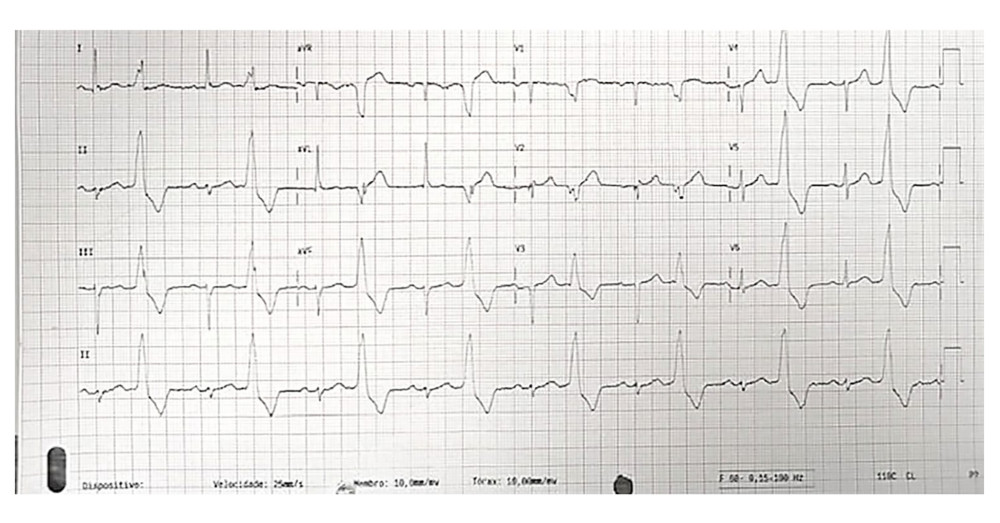 Figure 1. Emergency department 12-lead electrocardiogram (ECG). The 12-lead ECG shows sinus rhythm with frequent premature ventricular complexes (PVCs). The PVCs have left bundle branch block-like morphology, inferior axis, and early precordial transition, with an R/S ratio greater than 1 in lead V2.
 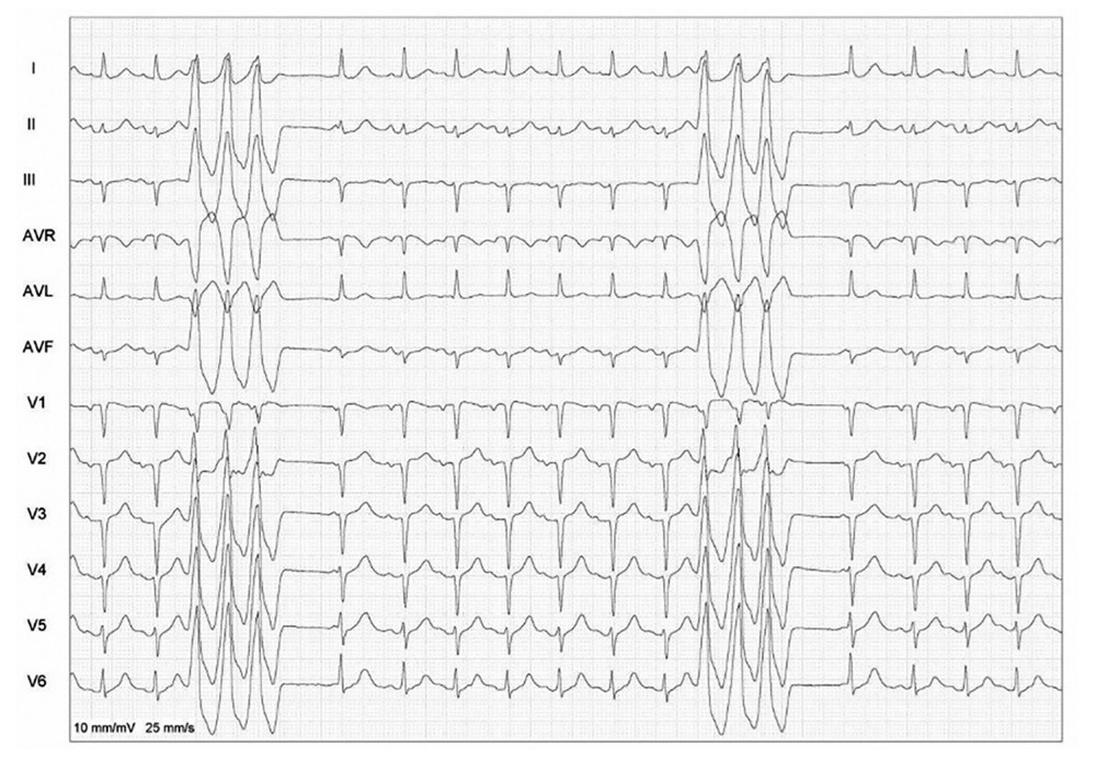 Figure 2. Intensive care unit electrocardiogram (ECG). The ECG shows nonsustained monomorphic ventricular tachycardia (NSVT) with the same QRS morphology as the premature ventricular complexes seen on the admission ECG, supporting a shared arrhythmogenic focus. Limb-lead analysis shows higher R-wave amplitude in lead II than in lead III and absence of septal q waves in leads I, II, and III. These findings support an anterosuperior left ventricular epicardial origin and are suggestive of left ventricular summit ventricular tachycardia.
 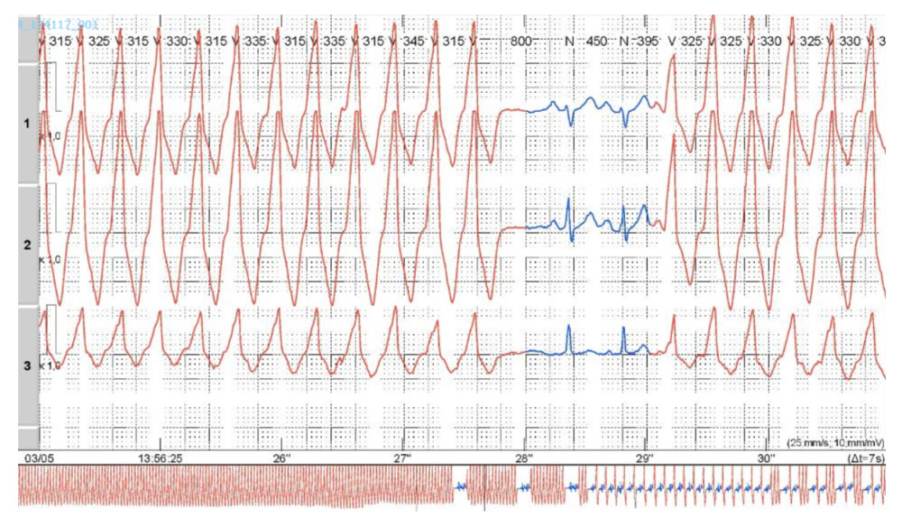 Figure 3. Twenty-four-hour Holter monitoring. The 24-hour Holter monitoring demonstrated a ventricular ectopic burden of approximately 65%, with frequent monomorphic premature ventricular complexes and recurrent sustained and nonsustained ventricular tachycardia, all sharing the same morphology as the index electrocardiogram.
 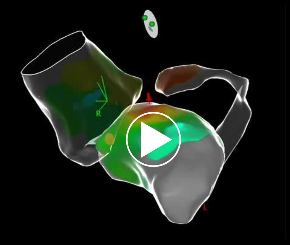 Video 1. Electrophysiological mapping. Activation mapping demonstrates the earliest ventricular activation in the region corresponding to the left ventricular summit, at a safe distance from the coronary arteries.
 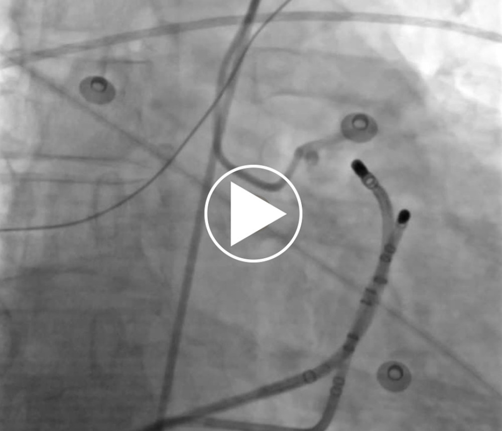 Video 2. Ablation strategy. Stepwise radiofrequency ablation from anatomically adjacent sites resulting in immediate and sustained suppression of ventricular arrhythmias.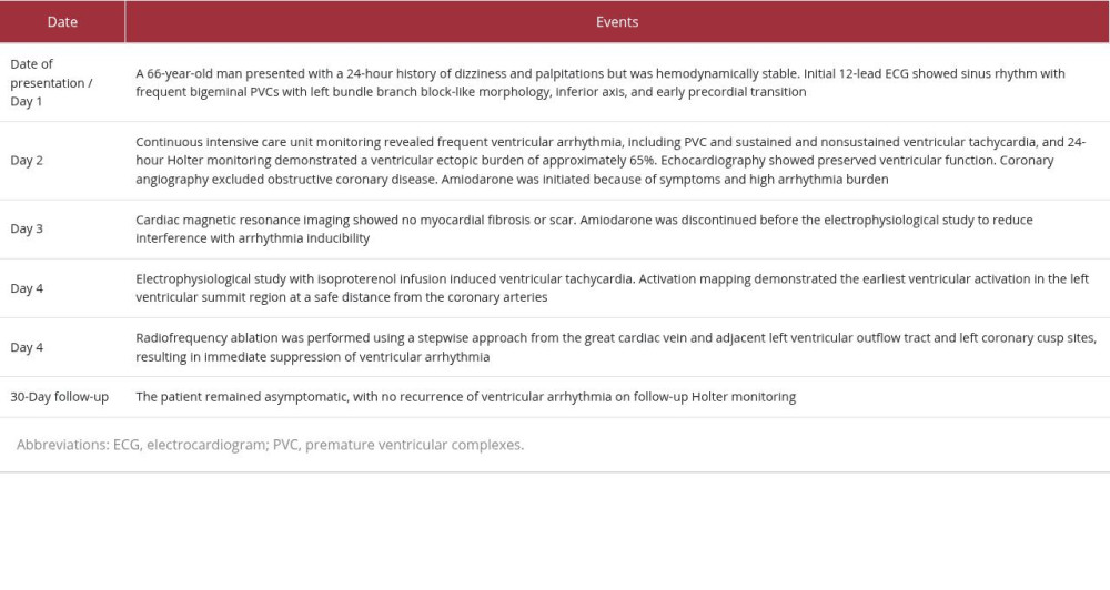
References
1. Das SK, Hawson J, Koh Y, Left ventricular summit arrhythmias: State-of-the-art review of anatomy, mapping, and ablation strategies: JACC Clin Electrophysiol, 2024; 10(11); 2516-39
2. Yamada T, McElderry HT, Doppalapudi H, Idiopathic ventricular arrhythmias originating from the left ventricular summit: Anatomic concepts relevant to ablation: Circ Arrhythm Electrophysiol, 2010; 3(6); 616-23
3. Yamada T, Yoshida N, Litovsky SH, Challenging radiofrequency catheter ablation of idiopathic ventricular arrhythmias originating from the left ventricular summit near the left main coronary artery: Circ Arrhythm Electrophysiol, 2016; 9(10); e004202
4. Anderson RD, Kumar S, Parameswaran R, Differentiating right- and left-sided outflow tract ventricular arrhythmias: Classical ECG signatures and prediction algorithms: Circ Arrhythm Electrophysiol, 2019; 12(6); e007392
5. Yakubov A, Salayev O, Hamrayev R, Sultankhonov S, A case of successful ablation of ventricular tachycardia focus in the left ventricular summit through the left atrial appendage: A case report: Eur Heart J Case Rep, 2018; 2(4); yty110
6. Shen B, Hu WM, Shao JM, Ventricular arrhythmias originating from different portions of the communicating vein of the left ventricular summit: Electrocardiographic characteristics and catheter ablation: BMC Cardiovasc Disord, 2024; 24(1); 421
Figures
Figure 1. Emergency department 12-lead electrocardiogram (ECG). The 12-lead ECG shows sinus rhythm with frequent premature ventricular complexes (PVCs). The PVCs have left bundle branch block-like morphology, inferior axis, and early precordial transition, with an R/S ratio greater than 1 in lead V2.Figure 2. Intensive care unit electrocardiogram (ECG). The ECG shows nonsustained monomorphic ventricular tachycardia (NSVT) with the same QRS morphology as the premature ventricular complexes seen on the admission ECG, supporting a shared arrhythmogenic focus. Limb-lead analysis shows higher R-wave amplitude in lead II than in lead III and absence of septal q waves in leads I, II, and III. These findings support an anterosuperior left ventricular epicardial origin and are suggestive of left ventricular summit ventricular tachycardia.Figure 3. Twenty-four-hour Holter monitoring. The 24-hour Holter monitoring demonstrated a ventricular ectopic burden of approximately 65%, with frequent monomorphic premature ventricular complexes and recurrent sustained and nonsustained ventricular tachycardia, all sharing the same morphology as the index electrocardiogram.Video 1. Electrophysiological mapping. Activation mapping demonstrates the earliest ventricular activation in the region corresponding to the left ventricular summit, at a safe distance from the coronary arteries.Video 2. Ablation strategy. Stepwise radiofrequency ablation from anatomically adjacent sites resulting in immediate and sustained suppression of ventricular arrhythmias. In Press
Case report 
Am J Case Rep In Press; DOI: 10.12659/AJCR.953543
Case report 
Am J Case Rep In Press; DOI: 10.12659/AJCR.954216
Case report 
Am J Case Rep In Press; DOI: 10.12659/AJCR.954932
Case report 
Am J Case Rep In Press; DOI: 10.12659/AJCR.954146
Most Viewed Current Articles
07 Dec 2021 : Case report
28,712,695
DOI :10.12659/AJCR.934347
Am J Case Rep 2021; 22:e934347
06 Dec 2021 : Case report
180,721
DOI :10.12659/AJCR.934406
Am J Case Rep 2021; 22:e934406
21 Jun 2024 : Case report
124,419
DOI :10.12659/AJCR.944371
Am J Case Rep 2024; 25:e944371
08 May 2026 : Case report
95,462
DOI :10.12659/AJCR.952158
Am J Case Rep 2026; 27:e952158