


09 September 2026: Articles 
Multidisciplinary Collaborative Treatment of Mediastinal Castleman Disease: A Case Report
Challenging differential diagnosis, Unusual or unexpected effect of treatment, Rare disease, Educational Purpose (only if useful for a systematic review or synthesis)
Yina Lang BCDEF 1, Jianchang Yao AG 2*
DOI: 10.12659/AJCR.953364
Am J Case Rep 2026; 27:e953364


Abstract
BACKGROUND: Castleman disease (CD) is a rare lymphoproliferative disorder, typically classified into unicentric Castleman disease (UCD) and multicentric Castleman disease (MCD). Except for painless masses, UCD usually presents no symptoms or only compression symptoms of surrounding tissues, while MCD presents as non-specific systemic symptoms such as fever, night sweats, fatigue, anorexia, weight loss, and hepatomegaly. Consequently, the diagnosis of CD relies on excision biopsy, making its diagnosis particularly challenging.
CASE REPORT: A 28-year-old man presenting with cough and hemoptysis was admitted to Deqing People’s Hospital. Chest computed tomography (CT) revealed a mediastinal mass below the tracheal carina, and puncture biopsy of the mass under bronchoscopy indicated lymphoid tissue hyperplasia. After multidisciplinary consultation, the possibility of malignancy in the mass was ruled out, surgical intervention was recommended, and an optimal surgical strategy was formulated. Ultimately, the patient underwent thoracoscopic-assisted mediastinal mass resection surgery and was diagnosed with UCD (hyaline-vascular variant) through postoperative histopathological examination and recovered well.
CONCLUSIONS: In this report, obtaining tissue samples through minimally invasive methods such as bronchoscopy facilitated the clarification of their pathological characteristics prior to surgery. Multidisciplinary team (MDT) meetings facilitated the development of optimal diagnostic and therapeutic strategies for complex diseases. For mediastinal diseases with unknown nature and non-specific clinical symptoms, performing bronchoscopic tissue biopsy and MDT before deciding whether to undergo surgery is beneficial for the patient in this report. This diagnostic and treatment process may be suitable for similar patients.
Keywords: Case Reports, Castleman disease, Surgery Department, Hospital
Introduction
Castleman disease (CD) is a rare lymphoproliferative disorder with an estimated incidence of less than 1/100 000 population [1,2]. CD manifests as localized lymphadenopathy, characterized by increased number of lymphoid follicles with degeneration of germinal centers and significant capillary proliferation, including the proliferation of follicular and interfollicular epithelium [1]. CD can occur in any part of the lymphatic system, with the mediastinum being the most common site of involvement, followed by the neck, abdomen, pelvis, and axilla [3–5].
Basing on the number of affected lymph nodes, CD is classified as unicentric or multicentric [4,6]. Unicentric Castleman disease (UCD) refers to a condition confined to a single anatomical site. In contrast, multicentric Castleman disease (MCD) involves multiple lymph nodes. UCD typically occurs in individuals aged 30 to 40 years, with no sex predilection, whereas MCD generally develops around the age of 60, with a slightly higher incidence in males than in females [1]. Histologically, CD is divided into hyaline-vascular, plasma cell type, and mixed variant [6]. The hyaline-vascular variant is commonly observed in UCD, characterized by large lymphoid follicles, prominent vascularization with hyalinization, and concentric layers of lymphocytes. Characteristic features include the “onion layer” appearance of mantle zone lymphocytes and a single penetrating central vessel [7]. In contrast, the plasma cell variant predominantly occurs in MCD, which is defined by a significant proliferation of polyclonal plasma cells in the interfollicular regions [8,9].
This report presents the diagnosis and treatment process of a case of mediastinal UCD. We attempted to obtain tissue through minimally invasive methods such as bronchoscopy to clarify the pathological properties and develop appropriate treatment plans through multidisciplinary team (MDT) meetings to avoid blind surgery. Ultimately, the patient achieved favorable therapeutic outcomes. We hope that our report can provide valuable references for the diagnosis and treatment of similar diseases.
Case Report
In late November 2025 a 28-year-old man with a history of cough and hemoptysis for 3 days sought medical assistance at the Respiratory Medicine outpatient department of Deqing People’s Hospital. Prior to this, the patient had no medical history. A chest computed tomography (CT) scan showed a round soft-tissue shadow below the tracheal carina and minor inflammatory fibrotic foci in both lungs. Considering the potential presence of complex diseases or malignant tumors, he was admitted to the Respiratory Medicine ward for further diagnosis and treatment.
After admission, physical examinations were performed and the results revealed: temperature 36.6 °C (normal range 36.3–37.2 °C), pulse rate 117 beats/minute (normal range 60–100 beats/minute), respiratory rate 17 breaths/minute (normal range 12–18 breaths/minute), and blood pressure 144/98 mm Hg (normal range 90-140/60-90 mm Hg). He had mildly increased breath sounds but without any dry or wet rales in both lungs, and other physical examination results were normal.
The abnormal results of laboratory tests conducted after admission were: serum alanine aminotransferase (ALT) 62 U/L (normal range 9–50 U/L), serum triglyceride (TG) 7.55 mmol/L (normal range 0–1.70 mmol/L), total cholesterol (TC) 7.43 mmol/L (normal range 0–5.20 mmol/L), blood uric acid (BUA) 604 μmol/L (normal range 202–416 μmol/L), and neuron-specific enolase (NSE) 97.10 μg/L (normal range, 0–30.10 μg/L).
Cervical color Doppler ultrasound revealed bilateral thyroid nodules (C-TIRADS Class 2) without cervical lymph node enlargement. Abdominal color Doppler ultrasound revealed hepatic adipose infiltration without abdominal lymph node enlargement. Enhanced CT of the chest showed a massive (53 × 43 mm) lesion in the mediastinum below the tracheal carina, with an average CT value of 44 Hu. Progressive and uniform enhancement was observed during contrast agent administration, with a CT value of 100 Hu (Figure 1A). Results of pulmonary ventilation function test, bronchodilation test, and 24-hour Holter ECG were all normal.
Considering the history of cough and hemoptysis over the past 3 days, the patient was administered tranexamic acid intravenously for hemostasis but no antibiotics due to the absence of definitive evidence of infection. Bronchoscopy and puncture biopsy of the mediastinal mass under bronchoscopy was performed, revealing mucous hyperemia of the right main bronchus with localized fine granular elevations (Figure 2). The pathological findings of the biopsy showed deep staining of cells, suggesting lymphoid tissue hyperplasia without lymphoma or other malignant tumors.
Based on the aforementioned examination results, a multidisciplinary consultation involving the Departments of Respiratory Medicine, Thoracic Surgery, Medical Oncology, Pathology, and Radiology was conducted, ultimately determining the necessity for surgical resection of the lesion. Subsequently, the patient was transferred to the Department of Thoracic Surgery for surgery. In early December 2025, he underwent thoracoscopic mediastinal tumor resection in the left lateral decubitus position with 45° forward tilt. The thoracoscopic lens was inserted at the fifth intercostal space along the midaxillary line for visualization. During the surgery, a mass measuring approximately 5 × 5 cm was observed in the mediastinum below the tracheal carina, closely associated with the right lower lobe of the lung and invading the vagus nerve. Therefore, partial resection of the right lower lung and vagotomy were performed to facilitate the successful removal of the mass. During the process of vagus nerve disconnection, a non-energy instrument was used to perform blunt/sharp separation of the vagus nerve. After the nerve was clearly exposed, an energy device was used to remove the surrounding tissue 3 mm from the nerve. Finally, the non-energy instrument was used again to transect nerves and ligate the residual ends. The surgery was successfully completed and a thoracic drainage tube was placed.
The resected mass had well-defined margins and homogeneous internal consistency (Figure 3), and the pathological examination revealed giant lymph node hyperplasia, consistent with Castleman disease (hyaline-vascular variant) (Figure 4). Immunohistochemical findings were: CD3 (partially positive), CD20 (partially positive), CK (negative), P40 (negative), CD21 (FDC+), Ki67 (+, 10%), CD31 (vessel-associated), Kappa (scattered positive), Lambda (scattered positive), and MUM1 (scattered positive). Based on these results, he was diagnosed with UCD (hyaline-vascular variant), recovered smoothly postoperatively, and was discharged 1 week later. The postoperative chest CT revealed complete resection of the mass (Figure 1B). In early January 2026 he was without any discomfort when he returned to the outpatient clinic for a follow-up chest CT scan, which showed good recovery (Figure 1C).
Discussion
Castleman disease was first described by Benjamin Castleman in 1954 and is typically divided into UCD and MCD [10,11]. UCD presents as a solitary, painless, non-malignant, and slow-growing mass localized to a single anatomical site, presenting with asymptomatic or compressive symptoms on adjacent structures as it enlarges [12]. MCD manifests with systemic symptoms such as fever, night sweats, fatigue, anorexia, weight loss, and hepatomegaly [13]. Therapeutically, surgical resection is the optimum selection for Guidewheel monoclonal Antibody-Based Therapeutic Regimens and is currently the preferred treatment option for MCD [14,15].
In clinical practice, the diagnosis of CD is difficult due to the lack of specific symptoms. According to the guideline of National Comprehensive Cancer Network (NCCN), the gold standard for diagnosis in CD is excision biopsy. However, the location of lesions in CD is not fixed, which poses diagnostic difficulties for patients with involvement of deep lymph nodes such as mediastinal lymph nodes. Currently, broader systemic endocrine, metabolic, and biochemical monitoring pathways are increasingly intersecting with modern surgical and diagnostic algorithms [16]. Interleukin-6 (IL-6) is a key driver of disease progression in some patients with CD [1]; therefore, monitoring its levels can be used to evaluate the efficacy of surgical or non-surgical treatment regimens in these patients.
For patients presenting with chest symptoms, X-ray examination is the preferred diagnostic modality, as it can reveal round, solitary masses in the thoracic region, including the lungs, hilar area, or paravertebral space. In chest CT, the vascular type of UCD typically appears as an isolated, well-demarcated, and localized nodular mass with a density equivalent to or lower than that of musculoskeletal density, and manifests as highly vascularized with homogeneous strong contrast enhancement on enhanced scans [17]. Similar diseases within the mediastinum, thymoma, and lymphoma have significantly weaker enhancement [18,19]. However, the excision biopsy of chest lesions require surgery, which seems hasty until malignant diseases such as lymphoma are ruled out. As shown in this report, obtaining partial tissue biopsy under bronchoscopy to determine whether surgery should be performed may be a good option. Although the pathological results of the mediastinal mass biopsy obtained via bronchoscopy failed to definitively confirm the disease in this report, the possibility of malignant lesions such as lymphoma was ruled out, which is beneficial for developing follow-up treatment plans. Therefore, performing a routine pathological biopsy preoperatively can be useful, as in reports by Phan et al, Chen et al, Ahmad et al, and Garcia et al [6,20–22].
Tumor diseases often present clinicians with complex situations. Over the past few decades, the literature has established MDT meetings as the standard diagnostic and treatment model for developing optimal treatment plans. In fact, experts with different backgrounds, skills, and clinical experience working together can develop the optimal treatment and diagnostic pathway [23]. In this report, MDT was conducted to discuss the patient’s subsequent treatment plan after completing the mediastinal lymph node biopsy under bronchoscopy. At the MDT meeting, we discussed histological data with pathologists and oncologists. All 3 parties evaluated the reliability of the specimens and unanimously agreed that tissue biopsy ruled out malignant diseases such as lymphoma. Experts from Thoracic Surgery and Radiology discussed the location of the lesion and the surrounding anatomical structures. Ultimately, thoracoscopic-assisted mediastinal mass resection surgery was chosen, resulting in complete recovery.
For mediastinal diseases with unknown nature and non-specific clinical symptoms, performing bronchoscopic tissue biopsy and MDT before deciding whether to undergo surgery can be beneficial for the patient, and this diagnostic and treatment process may be suitable for similar patients.
Conclusions
In summary, a case of mediastinal UCD was successfully diagnosed and treated in this report. Prior to the surgery, the possibility of malignant disease was ruled out through bronchoscopic mediastinal biopsy, and an appropriate surgical plan was developed through MDT. UCD needs to be considered in the differential diagnosis of a well-circumscribed enhancing mediastinal mass. Although the final diagnosis still relies on pathology results, preoperative biopsy can help rule out certain other possible diagnoses to guide subsequent treatment. For mediastinal diseases similar to the present case, this diagnostic and treatment process may be suitable.
Figures
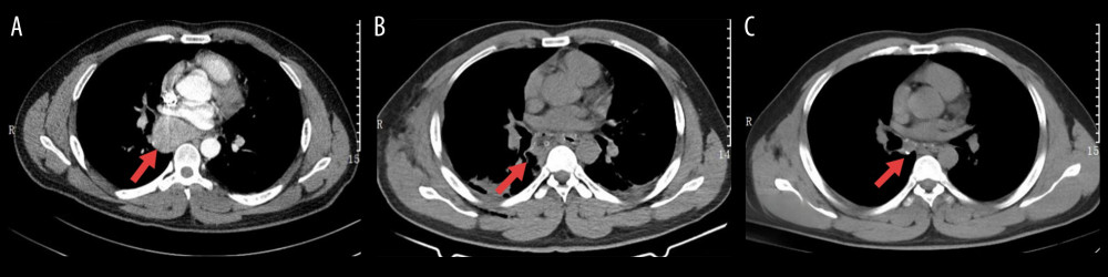 Figure 1. (A) Preoperative chest enhanced computed tomography revealed that the lesion was located below the tracheal carina and adjacent to the right main bronchus. (B) Postoperative chest computed tomography revealed complete resection of the mass. (C) Postoperative chest computed tomography scan 1 month later showed good recovery.
 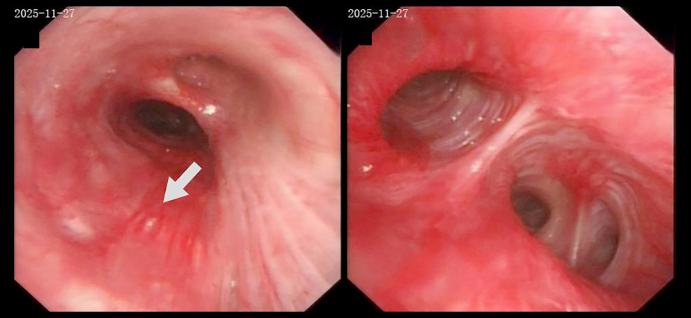 Figure 2. Bronchoscopy revealed mucosal congestion and swelling near the right main bronchial carina. Puncture biopsy of the mediastinal mass under bronchoscopy was performed at the site indicated by the arrow.
 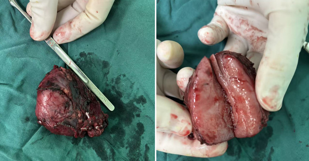 Figure 3. The resected mass had well-defined margins and homogeneous internal consistency.
 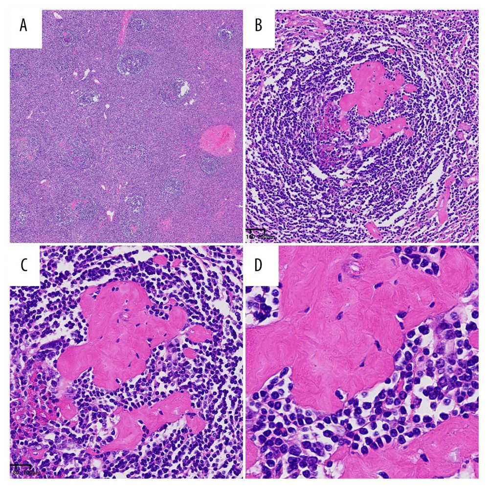 Figure 4. The pathological features of the patient’s lesion align with the histological characteristics of Castleman disease (hematoxylin-eosin stain). In the interlayered zone, lymphocytes are arranged in concentric circles resembling the layers of an onion, accompanied by penetration of transparent blood vessels through the atrophic germinal center. (A) Low-power view (hematoxylin-eosin, × 25), (B) High-power view (hematoxylin-eosin, × 200), (C) High-power view (hematoxylin-eosin, × 400), (D) High-power view (hematoxylin-eosin, × 800).
 References
1. Dispenzieri A, Fajgenbaum DC, Overview of Castleman disease: Blood, 2020; 135(16); 1353-64
2. Dégot T, Métivier A, Casnedi SThoracic manifestations of Castleman’s disease: Rev Pneumol Clin, 2009; 65(2); 101-7
3. McCarty MJ, Vukelja SJ, Banks PM, Angiofollicular lymph node hyperplasia (Castleman’s disease): Cancer Treat Rev, 1995; 21(4); 291-310
4. McAdams HP, Rosado-de-Christenson M, Fishback NF, Castleman disease of the thorax: Radiologic features with clinical and histopathologic correlation: Radiology, 1998; 209(1); 221-28
5. Jiang P, Huang Z, Liu W, Castleman disease presenting in the neck: A report of 3 cases and a literature review: Am J Case Rep, 2024; 25; e943214
6. Phan TT, Dong MH, Phan TP, A challenging case of angioproliferative Castleman’s disease in the mediastinum: Respir Med Case Rep, 2025; 58; 102287
7. Cronin DM, Warnke RA, Castleman disease: An update on classification and the spectrum of associated lesions: Adv Anat Pathol, 2009; 16(4); 236-46
8. Casper C, The aetiology and management of Castleman disease at 50 years: Translating pathophysiology to patient care: Br J Haematol, 2005; 129(1); 3-17
9. Cunha R, Silva R, Nogueira C, Unexpected diagnosis of concomitant Castleman’s disease following carotid body tumour resection: a case report and management review: EJVES Vasc Forum, 2025; 64; 75-77
10. Castleman B, Iverson L, Menendez VP, Localized mediastinal lymphnode hyperplasia resembling thymoma: Cancer, 1956; 9(4); 822-30
11. Haro A, Kuramitsu E, Fukuyama Y, Complete resection of unicentric Castleman disease in the superior mediastinum: A case report: Int J Surg Case Rep, 2016; 25; 44-47
12. Wang S, Chen S, Xu J, Cai S, Clinicopathological characteristics of unicentric retroperitoneal Castleman’s disease: A study of 14 cases: World J Surg Oncol, 2016; 14(1); 3
13. Yoshizaki K, Matsuda T, Nishimoto N, Pathogenic significance of interleukin-6 (IL-6/BSF-2) in Castleman’s disease: Blood, 1989; 74(4); 1360-67
14. Kligerman SJ, Auerbach A, Franks TJ, Castleman disease of the thorax: cCinical, radiologic, and pathologic correlation: From the radiologic pathology archives: Radiographics, 2016; 36(5); 1309-32
15. Zhang MY, Jia MN, Chen J, UCD with MCD-like inflammatory state: Surgical excision is highly effective: Blood Adv, 2021; 5(1); 122-28
16. Şahin Ç, Aydoğdu YF, Büyükkasap Ç, Investigation of homocysteine level after bariatric metabolic surgery, effect on vitamin B12 and folate levels: BMC Endocr Disord, 2024; 24(1); 237
17. Wong RSM, Unicentric Castleman disease: Hematol Oncol Clin North Am, 2018; 32(1); 65-73
18. Marx A, Pfister F, Schalke B, The different roles of the thymus in the pathogenesis of the various myasthenia gravis subtypes: Autoimmun Rev, 2013; 12(9); 875-84
19. Luan Y, Li X, Luan Y, Therapeutic challenges in peripheral T-cell lymphoma: Molecular Cancer, 2024; 23(1); 2
20. Chen HY, Wu SH, Chew FY, Mediastinal Castleman disease presenting as a paraspinal mass causing back pain and shortness of breath in a young adult: Heliyon, 2024; 10(24); e40792
21. Ahmad S, Iqbal SM, Qazi R, Isolated hilar mass in unicentric Castleman disease: Surgery as the definitive diagnosis and treatment: Cureus, 2025; 17(9); e92649
22. Garcia S, Fan J, Castleman disease presenting as a mediastinal mass: Cureus, 2021; 13(9); e18409
23. Tacchi G, Frasca L, Longo F, Crucitti P, Importance of the multidisciplinary approach in the surgical treatment of giant mediastinal neoplasms: A case series: AME Case Rep, 2024; 9; 33
Figures
Figure 1. (A) Preoperative chest enhanced computed tomography revealed that the lesion was located below the tracheal carina and adjacent to the right main bronchus. (B) Postoperative chest computed tomography revealed complete resection of the mass. (C) Postoperative chest computed tomography scan 1 month later showed good recovery.Figure 2. Bronchoscopy revealed mucosal congestion and swelling near the right main bronchial carina. Puncture biopsy of the mediastinal mass under bronchoscopy was performed at the site indicated by the arrow.Figure 3. The resected mass had well-defined margins and homogeneous internal consistency.Figure 4. The pathological features of the patient’s lesion align with the histological characteristics of Castleman disease (hematoxylin-eosin stain). In the interlayered zone, lymphocytes are arranged in concentric circles resembling the layers of an onion, accompanied by penetration of transparent blood vessels through the atrophic germinal center. (A) Low-power view (hematoxylin-eosin, × 25), (B) High-power view (hematoxylin-eosin, × 200), (C) High-power view (hematoxylin-eosin, × 400), (D) High-power view (hematoxylin-eosin, × 800). In Press
Case report 
Am J Case Rep In Press; DOI: 10.12659/AJCR.953543
Case report 
Am J Case Rep In Press; DOI: 10.12659/AJCR.954216
Case report
Am J Case Rep In Press; DOI: 10.12659/AJCR.954932
Case report 
Am J Case Rep In Press; DOI: 10.12659/AJCR.954146
Most Viewed Current Articles
07 Dec 2021 : Case report
28,712,695
DOI :10.12659/AJCR.934347
Am J Case Rep 2021; 22:e934347
06 Dec 2021 : Case report  180,721
DOI :10.12659/AJCR.934406
Am J Case Rep 2021; 22:e934406
21 Jun 2024 : Case report
124,419
DOI :10.12659/AJCR.944371
Am J Case Rep 2024; 25:e944371
08 May 2026 : Case report
95,462
DOI :10.12659/AJCR.952158
Am J Case Rep 2026; 27:e952158