


18 September 2026: Articles 
Prolonged Respiratory Failure With Pulmonary Fibroproliferative Complications Following Severe Drowning-Associated ARDS: A Case Report
Unusual clinical course
Katsutoshi Saito ABDE 1, Yoshihiro Miyake ADEF 1, Tomohiro Abe ABDE 1,2*, Hidenobu Ochiai G 1
DOI: 10.12659/AJCR.953531
Am J Case Rep 2026; 27:e953531


Abstract
BACKGROUND: Drowning-associated acute respiratory distress syndrome (ARDS) is generally considered a reversible condition, with most patients requiring only short-term mechanical ventilation. However, prolonged respiratory failure and fibroproliferative changes have rarely been described. We report a case of severe, prolonged ARDS following drowning complicated by recurrent pneumothorax and late-phase lung injury.
CASE REPORT: A 15-year-old girl developed ARDS following a drowning accident. Oxygenation collapsed shortly (PaO2/FiO2 ratio of 54.4), necessitating advanced ventilation and prone positioning. The clinical course was complicated by mediastinal emphysema and recurrent bilateral pneumothorax, associated with highly organized and cystic changes, requiring multiple chest tube insertions until day 57. Intensive treatment, including steroid pulses and nitric oxide inhalation, gradually improved respiratory function, resulting in the successful discontinuation of mechanical ventilation on day 53. Chest computed tomography on day 81 demonstrated marked improvement and resolution of organizing changes, with only limited residual organization. The patient was discharged home on day 93.
CONCLUSIONS: This case highlights that drowning-associated ARDS can, in rare cases, follow a prolonged course with fibroproliferative changes and recurrent complications such as pneumothorax. Nevertheless, even prolonged and complicated respiratory failure following drowning-associated ARDS may still demonstrate partial reversibility over time, suggesting the importance of avoiding premature therapeutic nihilism in similar cases.
Keywords: Drowning, Fibrosis, Pneumothorax, Respiratory Distress Syndrome
Introduction
Drowning causes approximately 300 000 deaths worldwide each year and is one of the leading causes of accidental death, particularly among children and young adults [1]. Following drowning, acute respiratory distress syndrome (ARDS) can develop as a result of surfactant dysfunction due to aspiration and subsequent inflammatory responses [2].
Clinical outcomes after drowning are strongly influenced by the presence and duration of cardiac arrest, which remains one of the most important determinants of mortality and neurological prognosis [3]. Nevertheless, respiratory dysfunction after drowning is generally transient and reversible, with most patients requiring only short-term mechanical ventilation, both in patients without cardiac arrest and in survivors of drowning-associated cardiac arrest [3]. However, knowledge regarding prolonged respiratory failure in drowning-associated ARDS remains limited, particularly regarding whether severe and prolonged fibroproliferative lung injury following drowning can result in persistent respiratory dysfunction. Here, we report the case of an adolescent patient with drowning-associated ARDS who developed an atypically prolonged fibroproliferative course complicated by recurrent pneumothorax and persistent respiratory failure, but who ultimately achieved substantial radiological and functional recovery. Through this case, we discuss factors potentially associated with the development of lung injury, the prolonged recovery course, and the possible reversibility of severe fibroproliferative changes. This case highlights that recovery may still be possible even in prolonged and complicated respiratory failure following drowning-associated ARDS.
Case Report
A 15-year-old girl was rescued from the sea after being found unresponsive in the water. Bystanders initiated cardiopulmonary resuscitation and called for an ambulance. The patient vomited during chest compression. Upon contact by the ambulance crew, her pulsation was palpable on her radial artery, but obvious hypoxemia was observed, prompting the request of a physician-staffed helicopter emergency medical system. At the scene, upon the physician’s arrival, the patient exhibited severe agitation and an altered level of consciousness. Her vital signs included a blood pressure of 110/72 mm Hg, heart rate of 102 beats/min, SpO2 of 74% with assisted bag-valve-mask ventilation using oxygen at 10 L/min, and a respiratory rate of 36 breaths/min. Due to the hypoxemia, endotracheal intubation was performed. No obvious aspirated material could be suctioned through the endotracheal tube at the time of intubation. During helicopter transport, her SpO2 was maintained at 85% to 90% by using manual ventilation.
Upon arrival at the emergency department, her vital signs were as follows: body temperature 37.0 °C, blood pressure 145/85 mm Hg, heart rate 120 beats/min, and SpO2 64% (with FiO2: 1.0). Physical examination revealed no obvious trauma. Arterial blood gas showed mixed acidosis, with pH of 7.10, partial pressure of carbon dioxide of 47.0 mm Hg, partial pressure of oxygen of 67.2 mm Hg, bicarbonate level of 13.9 mmol/L, base excess of −15.6 mmol/L, and lactate level of 8.66 mmol/L. Chest X-ray showed diffuse infiltrates in both lungs and no pneumothorax, and computed tomography (CT) of the chest demonstrated diffuse infiltrates in the dorsal lung fields and diffuse ground-glass opacities in the ventral lung fields (Figure 1).
The patient was diagnosed with acute respiratory distress syndrome (ARDS) due to drowning and was admitted to the intensive care unit. The overall clinical course, including ventilatory strategies, oxygenation trends, and major treatment, is summarized in Figure 2. Initially, the patient was managed with pressure-controlled ventilation, However, due to rapid deterioration of oxygenation with a PaO2/FiO2 (P/F) ratio of 54.4, airway pressure release ventilation with FiO2 1.0 and P-high/low 30/0 and full prone positioning for 16 hours per day were introduced on the same day. Oxygenation gradually improved, and prone positioning was discontinued when weaning progressed to P-high 20 and FiO2 0.3. On day 4, the patient developed mediastinal emphysema. Despite the spontaneous recovery from mediastinal emphysema on day 7, her respiratory status worsened, with FiO2 0.8, and a P/F ratio of approximately 100 led to the re-initiation of prone positioning. On day 9, due to continued worsening of respiratory status and imaging findings, nitric oxide inhalation therapy was started. Additionally, bovine pulmonary surfactant of 240 mg was administered on day 11 and day 19, but no significant improvement in oxygenation was observed. On day 19, suspected drug-induced pneumonia led to steroid pulse therapy (methylprednisolone 1 g for 3 days), but respiration function and imaging showed no response. On day 25, surgical tracheostomy was performed. Subsequently, oxygenation gradually improved, resulting in weaning of mechanical ventilation and tapering of nitric oxide therapy. On day 32, a right pneumothorax was identified on the chest X-ray, and a chest tube was subsequently inserted. Although her respiratory function improved, resulting in the cessation of nitric oxide therapy on day 34, it deteriorated on day 35. Chest CT showed a right-sided pneumothorax associated with highly organized cystic lung changes (Figure 3). The pneumothoraxes did not respond adequately to a single chest tube and recurred; thus, each episode was treated by a few chest tubes until day 57. Following the recovery of her respiratory function, mechanical ventilation was discontinued on day 53.
Her subsequent course was uneventful. Chest CT on day 82 demonstrated marked improvement of organizing changes, accompanied by bronchial wall thickening and diffuse ground-glass opacities in most areas, although residual fibrotic changes persisted in the right middle lobe, lingular segment, and bilateral lung apices (Figure 3). Pulmonary function tests showed restrictive impairment, with a vital capacity of 43.3% predicted. The 6-minute walk distance was 300 m, compared with an expected length of 794 m. Residual restrictive ventilatory impairment was considered to reflect residual pulmonary fibrotic changes and possible intensive care unit–acquired weakness. Rehabilitation was therefore continued, and the patient was discharged home without supplemental oxygen on day 93.
Discussion
Drowning-associated ARDS is known to result from surfactant depletion due to aspiration, alveolar epithelial injury, and inflammatory responses [2]. The severity and prognosis of drowning-associated ARDS are strongly influenced by the presence and duration of cardiac arrest, which remains one of the most important determinants of mortality and neurological prognosis [3]. Nevertheless, accumulating evidence indicates that respiratory failure in patients without cardiac arrest with severe hypoxemia and in survivors of drowning-associated cardiac arrest is typically transient, with most patients requiring only short-term mechanical ventilation, reflecting the generally reversible nature of drowning-related lung injury [4,5]. In contrast, the present case was characterized by prolonged and severe ARDS, with bystander cardiopulmonary resuscitation performed, and an unclear cardiac arrest status prior to medical contact. During the clinical course, highly organized and cystic lung changes developed, accompanied by recurrent pneumothorax, suggesting progression to a late fibroproliferative phase of ARDS. This clinical trajectory clearly differs from the typical natural course of drowning-associated ARDS, in which early improvement is generally expected.
Of note, the clinical course in the present case could have been influenced by several additional factors, including ventilator-induced lung injury, infection, and possible drug-induced lung injury. These secondary factors may have contributed to the development and persistence of lung injury, resulting in highly organized and cystic lung changes as well as recurrent pneumothorax. Thus, although drowning-associated lung injury likely initiated the respiratory failure, the prolonged fibroproliferative course cannot be attributed solely to drowning itself and was influenced by other secondary factors. Nevertheless, despite these complicated clinical features, the patient ultimately demonstrated substantial radiological and functional recovery over time. This case illustrates that even prolonged fibroproliferative respiratory failure following severe drowning-associated ARDS may still retain some reversibility.
Furthermore, lung injury following ARDS—regardless of etiology, including sepsis, pneumonia, or aspiration—is known to demonstrate a degree of reversibility [6]. During recovery from ARDS, a transient fibroproliferative response characterized by fibroblast proliferation and extracellular matrix deposition can occur; however, in many cases, these changes represent part of the repair process and gradually resolve over time. Long-term follow-up studies of ARDS survivors have shown that while some patients exhibit residual fibrotic-like changes on imaging or restrictive ventilatory impairment, these abnormalities are often mild and may partially or completely improve [6].
Several limitations of this case report warrant mention. First, the present case involved a young patient, which may have influenced the recovery course. Second, the prolonged recovery course was likely influenced by mixed etiologies that contributed to the severity and persistence of lung injury. Finally, this report describes a single successful case, and publication bias toward favorable outcomes should also be considered. Nevertheless, as illustrated in the present case, even severe and prolonged respiratory failure complicated by fibroproliferative changes and recurrent pneumothorax following drowning-associated ARDS may still retain some reversibility. This case may therefore serve as a caution against premature therapeutic nihilism in similarly prolonged and complicated cases.
Conclusions
Prolonged respiratory failure can occur after drowning-associated ARDS. Although fibroproliferative changes were associated with recurrent complications, including pneumothorax, substantial recovery was ultimately achieved despite severe late-phase lung injury. The clinical course was likely influenced by multiple factors, including drowning-related lung injury, ventilator-induced lung injury, and medications administered during intensive care management. This case suggests that recovery may still be possible even in prolonged and complicated presentations of drowning-associated ARDS.
Figures
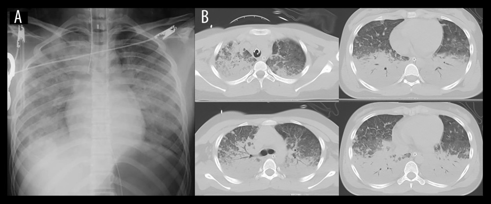 Figure 1. Chest imaging on admission(A) Chest X-ray demonstrated bilateral diffuse pulmonary infiltrates. (B) Chest computed tomography revealed diffuse bilateral ground-glass opacities and consolidation.
 ![Clinical course of respiratory managementThe timeline shows major interventions, ventilator modes, corticosteroid therapy, and respiratory parameters during hospitalization. The solid line represents the PaO2/FiO2 (P/F) ratio, and the dotted line indicates positive end-expiratory pressure (PEEP). Ventilator modes used during the course are shown at the bottom (airway pressure release ventilation [APRV] and pressure support ventilation [PSV]). Gray bars indicate the dose of methylprednisolone (mPSL). Arrows denote the timing of tracheostomy and chest tube insertion, and the horizontal bar represents the period of inhaled nitric oxide (NO) therapy. Severe hypoxemia initially required high PEEP and APRV support; oxygenation gradually improved over time, allowing reduction of ventilatory support.](https://jours.isi-science.com/imageXml.php?i=amjcaserep-27-e953531-g002.jpg&idArt=953531&w=1000) Figure 2. Clinical course of respiratory managementThe timeline shows major interventions, ventilator modes, corticosteroid therapy, and respiratory parameters during hospitalization. The solid line represents the PaO2/FiO2 (P/F) ratio, and the dotted line indicates positive end-expiratory pressure (PEEP). Ventilator modes used during the course are shown at the bottom (airway pressure release ventilation [APRV] and pressure support ventilation [PSV]). Gray bars indicate the dose of methylprednisolone (mPSL). Arrows denote the timing of tracheostomy and chest tube insertion, and the horizontal bar represents the period of inhaled nitric oxide (NO) therapy. Severe hypoxemia initially required high PEEP and APRV support; oxygenation gradually improved over time, allowing reduction of ventilatory support.
Figure 2. Clinical course of respiratory managementThe timeline shows major interventions, ventilator modes, corticosteroid therapy, and respiratory parameters during hospitalization. The solid line represents the PaO2/FiO2 (P/F) ratio, and the dotted line indicates positive end-expiratory pressure (PEEP). Ventilator modes used during the course are shown at the bottom (airway pressure release ventilation [APRV] and pressure support ventilation [PSV]). Gray bars indicate the dose of methylprednisolone (mPSL). Arrows denote the timing of tracheostomy and chest tube insertion, and the horizontal bar represents the period of inhaled nitric oxide (NO) therapy. Severe hypoxemia initially required high PEEP and APRV support; oxygenation gradually improved over time, allowing reduction of ventilatory support. 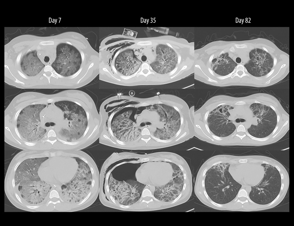 Figure 3. Serial chest computed tomography during the clinical courseChest computed tomography scans performed on days 7, 35, and 82 after admission show the temporal evolution of lung injury. On day 7, diffuse bilateral ground-glass opacities and consolidation compatible with acute respiratory distress syndrome were observed. On day 35, persistent bilateral infiltrates with fibroproliferative changes were evident, and a right-sided pneumothorax requiring chest tube placement had developed. By day 82, the lung opacities had markedly improved, with substantial resolution of the previously observed abnormalities.
 References
1. World Health Organization (Geneva): Drowning: World Health Organization, 2023 Available from: https://www.who.int/news-room/fact-sheets/detail/drowning
2. Bierens JJLM, Lunetta P, Tipton M, Warner DS, Physiology of drowning: A review: Physiology, 2016; 31; 147-66
3. Markarian T, Loundou A, Heyer V, Drowning classification: A reappraisal of clinical presentation and prognosis for severe cases: Chest, 2020; 158; 596-602
4. Son KL, Hwang SK, Choi HJ, Clinical features and prognostic factors in drowning children: A regional experience: Korean J Pediatr, 2016; 59; 212-17
5. Reizine F, Michelet P, Delbove A, Development and validation of a clinico-biological score to predict outcomes in patients with drowning-associated cardiac arrest: Am J Emerg Med, 2024; 81; 69-74
6. Herridge MS, Tansey CM, Matté A, Functional disability 5 years after acute respiratory distress syndrome: N Engl J Med, 2011; 364; 1293-304
Figures
Figure 1. Chest imaging on admission(A) Chest X-ray demonstrated bilateral diffuse pulmonary infiltrates. (B) Chest computed tomography revealed diffuse bilateral ground-glass opacities and consolidation.Figure 2. Clinical course of respiratory managementThe timeline shows major interventions, ventilator modes, corticosteroid therapy, and respiratory parameters during hospitalization. The solid line represents the PaO2/FiO2 (P/F) ratio, and the dotted line indicates positive end-expiratory pressure (PEEP). Ventilator modes used during the course are shown at the bottom (airway pressure release ventilation [APRV] and pressure support ventilation [PSV]). Gray bars indicate the dose of methylprednisolone (mPSL). Arrows denote the timing of tracheostomy and chest tube insertion, and the horizontal bar represents the period of inhaled nitric oxide (NO) therapy. Severe hypoxemia initially required high PEEP and APRV support; oxygenation gradually improved over time, allowing reduction of ventilatory support.Figure 3. Serial chest computed tomography during the clinical courseChest computed tomography scans performed on days 7, 35, and 82 after admission show the temporal evolution of lung injury. On day 7, diffuse bilateral ground-glass opacities and consolidation compatible with acute respiratory distress syndrome were observed. On day 35, persistent bilateral infiltrates with fibroproliferative changes were evident, and a right-sided pneumothorax requiring chest tube placement had developed. By day 82, the lung opacities had markedly improved, with substantial resolution of the previously observed abnormalities. In Press
Case report 
Am J Case Rep In Press; DOI: 10.12659/AJCR.953317
Case report 
Am J Case Rep In Press; DOI: 10.12659/AJCR.954643
Case report 
Am J Case Rep In Press; DOI: 10.12659/AJCR.954074
Case report 
Am J Case Rep In Press; DOI: 10.12659/AJCR.953592
Most Viewed Current Articles
07 Dec 2021 : Case report
28,712,429
DOI :10.12659/AJCR.934347
Am J Case Rep 2021; 22:e934347
06 Dec 2021 : Case report  180,530
DOI :10.12659/AJCR.934406
Am J Case Rep 2021; 22:e934406
21 Jun 2024 : Case report
124,276
DOI :10.12659/AJCR.944371
Am J Case Rep 2024; 25:e944371
08 May 2026 : Case report
95,045
DOI :10.12659/AJCR.952158
Am J Case Rep 2026; 27:e952158